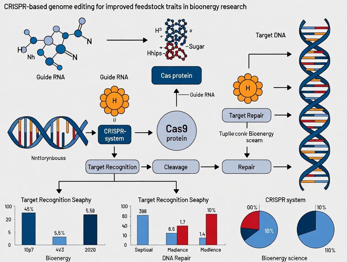
CRISPR Genome Editing: Revolutionizing Feedstock Traits for Enhanced Biomolecule Production
This article provides a comprehensive overview of CRISPR-based genome editing strategies specifically tailored to optimize microbial, plant, and mammalian feedstocks for biomedical and drug development applications.
CRISPR Genome Editing: Revolutionizing Feedstock Traits for Enhanced Biomolecule Production
Abstract
This article provides a comprehensive overview of CRISPR-based genome editing strategies specifically tailored to optimize microbial, plant, and mammalian feedstocks for biomedical and drug development applications. It explores foundational principles, methodological applications for key trait improvements, strategies to overcome technical hurdles, and validation frameworks for comparative analysis. Targeted at researchers and drug development professionals, this review synthesizes current advancements to guide the engineering of superior production platforms for therapeutics, vaccines, and other valuable biomolecules.
CRISPR 101: Core Principles and Feedstock Engineering Fundamentals
Within the broader thesis of developing CRISPR-based genome editing for improved feedstock traits—such as enhanced nutritional profiles, disease resistance, and abiotic stress tolerance in crops and livestock—understanding the transition from a prokaryotic immune system to a programmable laboratory tool is fundamental. This section provides critical notes on key CRISPR-Cas systems and their tailored applications in feedstock research.
Application Note 1: Choosing the Cas Nuclease for Feedstock Trait Engineering The selection of the Cas enzyme dictates the experimental strategy and potential outcomes. The table below summarizes the quantitative characteristics of the most commonly used nucleases.
Table 1: Comparison of Key CRISPR-Cas Nucleases for Genome Editing
| Nuclease | Origin | Guide RNA (gRNA) Length | PAM Sequence | Primary Cleavage Output | Key Application in Feedstock Research |
|---|---|---|---|---|---|
| SpCas9 | S. pyogenes | ~20 nt | 5'-NGG-3' | DSB | High-efficiency gene knockouts in plants (e.g., maize, soybean) and animal zygotes. |
| Cas12a (Cpf1) | Prevotella and Francisella | ~20-24 nt | 5'-TTTV-3' | DSB with staggered ends | Multiplexed editing in AT-rich genomic regions; suitable for polyploid crop species. |
| Cas9-Nickase (nCas9) | Engineered SpCas9 | ~20 nt | 5'-NGG-3' | Single-strand nick | Base editing when fused to deaminase; reduces off-target effects in elite livestock lines. |
| dCas9 | Catalytically dead SpCas9 | ~20 nt | 5'-NGG-3' | No cleavage | Transcriptional repression/activation (CRISPRi/a) of metabolic pathways without altering DNA sequence. |
Application Note 2: Delivery Methods for Feedstock Systems Efficient delivery remains a bottleneck. For plants, Agrobacterium-mediated transformation of embryonic tissue or RNP delivery via biolistics are standard. For livestock, microinjection of CRISPR components into zygotes or using viral vectors (e.g., lentivirus) in somatic cells is prevalent. The choice impacts editing efficiency, off-target rate, and regulatory status (GMO vs. non-transgenic).
Application Note 3: Verification and Screening Post-editing, a robust screening pipeline is required. This involves: 1) PCR amplification of the target locus, 2) Deep sequencing (amplicon-seq) to quantify editing efficiency and heterogeneity, and 3) For crops, regeneration of whole plants from edited calli and genotyping of T0/T1 generations to assess heritability.
Detailed Experimental Protocols
Protocol 1: Design and Validation of gRNAs for a Target Feedstock Gene Objective: To design and empirically test the in vitro cleavage efficiency of gRNAs targeting a gene of interest (e.g., a lignin biosynthesis gene in sorghum). Materials: See "The Scientist's Toolkit" below. Method: 1. Identification: Input the target gene sequence into a design tool (e.g., CHOPCHOP, Benchling). Select 3-5 gRNAs with high on-target and low off-target scores. 2. Cloning: Synthesize oligonucleotides for each gRNA, anneal, and ligate into a CRISPR expression plasmid (e.g., pBUN411) downstream of the U6 promoter. 3. In Vitro Transcription: Using the cloned plasmid as template, transcribe gRNA with a T7 polymerase kit. Purify using spin columns. 4. In Vitro Cleavage Assay: a. Amplify a ~500-800 bp genomic DNA fragment encompassing the target site from the feedstock organism. b. Set up a 20 µL reaction: 100 ng PCR product, 50 nM purified SpCas9 protein, 50 nM gRNA, 1X Cas9 reaction buffer. c. Incubate at 37°C for 1 hour. Terminate with Proteinase K. d. Run products on a 2% agarose gel. Successful cleavage yields two smaller fragments. Quantify efficiency using gel analysis software. Analysis: The gRNA yielding >80% cleavage in vitro is prioritized for in vivo experiments.
Protocol 2: Agrobacterium-Mediated CRISPR/Cas9 Delivery in a Model Plant (Tomato) Objective: To generate stable, heritable knockouts in a diploid crop species. Method: 1. Vector Assembly: Clone the validated gRNA expression cassette and a plant-codon-optimized Cas9 gene (driven by a 35S promoter) into a T-DNA binary vector with a plant selection marker (e.g., kanamycin resistance). 2. Transformation: Introduce the binary vector into Agrobacterium tumefaciens strain LBA4404 via electroporation. 3. Plant Transformation: a. Surface-sterilize tomato seeds, germinate on MS media, and use cotyledon explants. b. Immerse explants in the Agrobacterium suspension (OD600 = 0.5) for 10 minutes. c. Co-cultivate on MS media for 48 hours in the dark. d. Transfer explants to selection media (containing kanamycin and cefotaxime) to induce callus formation and shoot regeneration (4-6 weeks). 4. Regeneration and Genotyping: a. Transfer regenerated shoots to rooting media. b. Extract genomic DNA from leaf tissue of putative transgenic plantlets. c. Perform PCR on the target locus and sequence the products. Use TIDE or ICE analysis to quantify indel frequencies.
Diagrams & Visualizations
Title: Native CRISPR-Cas Adaptive Immunity Pathway
Title: CRISPR Genome Editing Workflow for Feedstock Research
The Scientist's Toolkit: Research Reagent Solutions
Table 2: Essential Materials for CRISPR-based Feedstock Genome Editing
| Reagent / Material | Supplier Examples | Function in Experiment |
|---|---|---|
| High-Fidelity DNA Polymerase (e.g., Q5) | NEB, Thermo Fisher | Accurate amplification of target genomic loci for gRNA testing and genotyping. |
| T7 RNA Polymerase Kit | NEB, Thermo Fisher | For in vitro transcription of gRNAs for validation assays or RNP complex formation. |
| Purified Recombinant Cas9 Protein | IDT, Thermo Fisher, in-house | For in vitro cleavage assays or direct delivery of RNP complexes, reducing off-target effects. |
| CRISPR-Cas9 Expression Vector (e.g., pBUN411) | Addgene | A plant-optimized binary vector for easy cloning of gRNAs and expression of Cas9. |
| Agrobacterium Strain LBA4404 | Various (CICC, lab stocks) | Standard disarmed strain for stable transformation of dicot plant species. |
| Plant Tissue Culture Media (MS Basal) | PhytoTech Labs, Duchefa | Provides essential nutrients for regeneration of whole plants from edited explants. |
| Next-Generation Sequencing Kit (Amplicon) | Illumina, Paragon Genomics | Enables deep sequencing of target loci to quantify editing efficiency and profile mutations. |
| Genomic DNA Extraction Kit (Plant/Animal) | Qiagen, Zymo Research | High-quality DNA extraction from tough feedstock tissues (e.g., leaf, muscle, seed). |
Within the broader thesis on CRISPR-based genome editing for improved feedstock traits, defining the ideal cellular host for therapeutic protein production is paramount. An ideal feedstock cell line must exhibit specific, engineerable traits to maximize yield, ensure product quality, and maintain economic viability. This application note details these key traits, supported by quantitative data, and provides protocols for their assessment and enhancement via genome editing.
Key Traits of an Ideal Feedstock: Quantitative Analysis
Table 1: Quantitative Targets for Ideal Feedstock Cell Lines
| Trait Category | Specific Parameter | Ideal Target / Benchmark | Measurement Method |
|---|---|---|---|
| Productivity | Specific Productivity (qP) | >50 pg/cell/day | ELISA / Metabolite analysis |
| Volumetric Titer | >5 g/L for mAbs | Product concentration assay | |
| Growth & Stability | Maximum Viable Cell Density (VCD) | >20 x 10^6 cells/mL | Automated cell counter |
| Integrated Viable Cell Density (IVCD) | High, process-dependent | Calculation from VCD over time | |
| Culture Longevity (Stationary Phase) | >7 days | Viability tracking | |
| Product Quality | Glycan Profile Consistency | >90% target glycoform (e.g., afucosylation) | HPAEC-PAD or LC-MS |
| Aggregate Formation | <5% | Size-exclusion chromatography (SEC) | |
| Metabolic Fitness | Lactate Metabolism Shift | Lactate production to consumption phase | Bioanalyzer / YSI analyzer |
| Ammonia Production | Low (<5 mM) | Biochemical assay | |
| Genetic Stability | Target Gene Expression Stability | <20% decrease over 60 generations | qPCR / Flow cytometry |
| Robustness | Resilience to Bioprocess Stress (pH, Osmolality) | High viability maintenance | Stress tests & viability assays |
Experimental Protocols
Protocol 2.1: CRISPR-Mediated Knock-In for Enhanced Specific Productivity Objective: Integrate a high-expression promoter upstream of the therapeutic gene locus to boost qP. Materials: CHO-K1 cells, Cas9 ribonucleoprotein (RNP), donor DNA template (ssODN with homology arms), Nucleofector Kit, growth media. Procedure:
- Design gRNA targeting a genomic "safe harbor" (e.g., AAVS1 locus in CHO cells).
- Form RNP complex: Incubate 10 µg Cas9 protein with 5 µg synthetic gRNA for 10 min at 25°C.
- Prepare donor template: 2 µg ssODN containing promoter-therapeutic gene cassette flanked by 80 bp homology arms.
- Harvest 1x10^6 log-phase cells, resuspend in Nucleofector solution with RNP and donor DNA.
- Electroporate using manufacturer's program.
- Recover cells in pre-warmed media for 48 hours before selection or single-cell cloning.
- Screen clones via junction PCR and quantify qP via fed-batch assay (Protocol 2.3).
Protocol 2.2: Assessment of Genetic Stability Using Long-Term Passage Objective: Determine the stability of CRISPR-edited traits over extended culture. Materials: Edited clonal cell line, seed train flasks, viability stain, genomic DNA extraction kit. Procedure:
- Initiate triplicate cultures of the edited clone at 0.3 x 10^6 cells/mL.
- Passage cells every 3-4 days, maintaining sub-confluent density. Count VCD and viability at each passage.
- At passages 5, 15, 30, 45, and 60, harvest 1x10^6 cells for gDNA extraction.
- Quantify target gene copy number via digital PCR relative to a reference gene.
- Correlate copy number with specific productivity (from periodic fed-batch assays) over generations.
Protocol 2.3: Micro-scale Fed-Batch Assay for Trait Phenotyping Objective: Characterize growth, metabolism, and productivity of engineered clones in a high-throughput format. Materials: 96-deep well plates, automated liquid handler, basal and feed media, metabolite analyzer. Procedure:
- Inoculate clones in 1 mL basal media at 0.3 x 10^6 cells/mL in 96-deep well plates.
- Incubate at 37°C, 5% CO2, 85% humidity with orbital shaking.
- On days 3, 5, and 7, add 0.15 mL of feed media.
- Daily, sample 50 µL from designated wells for VCD/viability (automated counter) and metabolite analysis (glucose, lactate, ammonia).
- On harvest day (day 10-14), centrifuge plates and collect supernatant for titer (ELISA) and product quality (SEC-HPLC) analysis.
Visualizing Pathways and Workflows
Diagram Title: CRISPR Workflow for Feedstock Engineering
Diagram Title: Key Traits Engineered by CRISPR for Ideal Feedstock
The Scientist's Toolkit: Research Reagent Solutions
Table 2: Essential Reagents for Feedstock Trait Engineering & Analysis
| Reagent / Material | Function / Application | Example Vendor/Code |
|---|---|---|
| CRISPR-Cas9 RNP Complex | Direct delivery of editing machinery; high efficiency, reduced off-target. | Synthego TrueCut Cas9 Protein + sgRNA |
| Single-Stranded Oligo Donor (ssODN) | Homology-directed repair template for precise knock-ins. | IDT Ultramer DNA Oligo |
| Cell Line Nucleofector Kit | High-efficiency transfection of hard-to-transfect feedstock cells (e.g., CHO). | Lonza Kit V, SF Cell Line 4D-Nucleofector X |
| CloneSelect Single-Cell Printer | Isolation of single cells for clonal derivation with high viability assurance. | Molecular Devices Firma |
| GlycoWorks RapiFluor-MS N-Glycan Kit | Rapid profiling of critical quality attribute (glycosylation). | Waters Corporation |
| Octet BLI Systems & Protein A Biosensors | Real-time, label-free titer measurement for high-throughput screening. | Sartorius |
| Live Cell Analysis Instrument (e.g., Incucyte) | Continuous monitoring of cell growth, confluence, and viability. | Sartorius Incucyte SX5 |
| Metabolite Bioanalyzer (e.g., Nova) | Automated measurement of glucose, lactate, and other key metabolites. | Nova Bioprofile FLEX2 |
Application Notes
Within the broader thesis on CRISPR-based genome editing for improved feedstock traits, engineering target organisms as production platforms is pivotal. These platforms are optimized to produce high-value compounds, from therapeutic proteins to industrial enzymes and biofortified crops. CRISPR technology enables precise, multiplexed edits to overcome bottlenecks in yield, quality, and scalability across kingdoms.
The following tables summarize key quantitative performance data for engineered platforms.
Table 1: CRISPR-Enhanced Microbial Platforms (E. coli and S. cerevisiae)
| Organism | Target Gene/Pathway | Edit Type | Output Metric | Result (vs. Wild-Type/Control) | Primary Product |
|---|---|---|---|---|---|
| E. coli | gallU, endA | Knockout | Plasmid Yield | 4.8-fold increase | Recombinant DNA |
| E. coli | T7 RNA Polymerase Locus | Integration | Protein Titer | 2.1 g/L (Fed-Batch) | scFv Antibody |
| S. cerevisiae | GRE3, ALD6 | Multiplex KO | Ethanol Yield | 11% increase | Biofuel (Ethanol) |
| S. cerevisiae | δ-Integration Sites | Multi-copy Integration | Protein Titer | 1.5 g/L (Shake Flask) | Human Serum Albumin |
Table 2: CRISPR-Engineered Mammalian Cell Platforms (CHO and HEK293)
| Cell Line | Target Locus | Edit Type | Output Metric | Result | Primary Product |
|---|---|---|---|---|---|
| CHO-S | FUT8 (α-1,6-fucosyltransferase) | Knockout | Afucosylated Antibody Proportion | >95% of pool | Monoclonal Antibody (mAb) |
| CHO-K1 | GS Locus | Site-Specific Integration | Stable Pool Titer | ~3 g/L (Batch) | IgG1 |
| HEK293T | AAVS1 Safe Harbor | Knock-in (Reporter) | Transfection Efficiency | ~85% GFP+ cells | Viral Vector Proteins |
Table 3: CRISPR-Improved Plant-Based Platforms (Nicotiana and Arabidopsis)
| Plant Species | Target Trait | Target Gene(s) | Edit Type | Quantitative Improvement | Application |
|---|---|---|---|---|---|
| Nicotiana benthamiana | Protein Yield | RNA-dependent RNA polymerase (RdRp) genes | Knockout | 2- to 3-fold increase in transient expression | Plant-made pharmaceuticals |
| Arabidopsis thaliana | Seed Oil Content | FAD2 | Knockout | Oleic acid increase from 20% to 60% | Nutritious Feedstock |
| Oryza sativa | Vitamin Precursor | LYC and CRTISO | Knock-in/Activation | β-carotene accumulation (Pro-Vitamin A) | Biofortified Crop |
Experimental Protocols
Protocol 1: Multiplexed Gene Knockout inS. cerevisiaefor Pathway Engineering
Objective: To simultaneously disrupt GRE3 (aldose reductase) and ALD6 (cytosolic aldehyde dehydrogenase) in yeast to reduce glycerol and acetate byproducts, redirecting carbon flux toward ethanol.
Materials: See "Research Reagent Solutions" below.
Method:
- gRNA Design & Cassette Assembly: Design two 20-nt guide RNA sequences targeting GRE3 and ALD6 using a validated online tool (e.g., CHOPCHOP). Clone gRNA sequences into the BsaI sites of plasmid pROS11 (expressing gRNA, SNR52 promoter, tRNA for processing).
- Repair Template Preparation: Synthesize two double-stranded DNA repair templates (~100 bp each) containing stop codons and frameshifts flanked by 50-bp homology arms to the target sites.
- Transformation: Co-transform S. cerevisiae strain BY4741 with the assembled gRNA plasmid and the two repair templates using the standard lithium acetate/PEG method.
- Selection & Screening: Plate on synthetic complete media lacking uracil to select for the gRNA plasmid. After 72h, patch colonies onto YPD plates. Screen for edits by colony PCR using primers flanking each target site.
- Validation: Sanger sequence PCR products. Confirm phenotypic change by performing small-scale fermentations in YP media with 20% glucose and measuring ethanol titers via HPLC.
Protocol 2:FUT8Knockout in CHO Cells for Afucosylated Antibody Production
Objective: Generate a clonal CHO cell line deficient in α-1,6-fucosyltransferase (FUT8) to enhance antibody-dependent cellular cytotoxicity (ADCC) of produced antibodies.
Method:
- RNP Complex Formation: Resuspend 60 pmol of chemically synthesized crRNA (targeting FUT8 exon 1) and tracrRNA in duplex buffer, heat to 95°C for 5 min, and cool. Complex with 40 pmol of purified SpCas9 protein to form ribonucleoprotein (RNP). Incubate 10 min at RT.
- CHO Cell Electroporation: Harvest log-phase CHO-S cells, wash with PBS. Resuspend 1e6 cells in 100 µL electroporation buffer (P3 Primary Cell Solution). Mix with RNP complex and transfer to a 100-µL electroporation cuvette. Electroporate using a 4D-Nucleofector (program CA-137).
- Recovery & Single-Cell Cloning: Immediately add pre-warmed medium, transfer to a plate. After 48h, begin puromycin selection (1 µg/mL) for 5 days. Recover cells, then seed by limiting dilution in 96-well plates for clonal isolation.
- Genotypic Screening: Extract genomic DNA from expanded clones. Perform PCR on the FUT8 target region and subject to T7 Endonuclease I assay. Sequence clones showing cleavage to confirm indels.
- Phenotypic Validation: Confirm FUT8 KO by lectin blotting with Aleuria aurantia lectin (AAL) on purified IgG. Measure ADCC activity using a reporter bioassay against target antigen-expressing cells.
Protocol 3:RdRpGene Knockout inNicotiana benthamianafor Enhanced Transient Expression
Objective: Generate stable N. benthamiana knockout lines defective in RNAi machinery to increase recombinant protein accumulation during Agrobacterium-mediated transient expression.
Method:
- CRISPR Construct Assembly: Using Golden Gate cloning, assemble a plant expression vector (e.g., pYLCRISPR/Cas9) with two gRNAs targeting conserved regions of essential RdRp1 and RdRp2 genes.
- Agrobacterium Transformation & Plant Transformation: Transform the assembled vector into A. tumefaciens strain GV3101. Transform wild-type N. benthamiana leaf discs via standard Agrobacterium co-cultivation. Regenerate plants on kanamycin-containing media.
- Regeneration & Genotyping: Transfer regenerated shoots (T0 plants) to rooting media. Screen by PCR on leaf tissue to confirm the presence of the Cas9 transgene. Sequence the target loci in PCR-positive plants to identify biallelic mutations.
- Homozygous Line Selection: Self-pollinate T0 plants with desired edits. Genotype T1 seedlings to identify homozygous, transgene-free lines (segregating out the Cas9 T-DNA).
- Functional Assay: Infiltrate leaves of wild-type and rdrp KO lines with Agrobacterium harboring a GFP expression vector. Image fluorescence at 3-5 days post-infiltration (dpi) and quantify total soluble protein and GFP yield by spectrophotometry and ELISA, respectively.
Visualizations
Title: CRISPR Redirects Yeast Carbon Flux to Ethanol
Title: Workflow for Generating FUT8-KO CHO Cell Line
Title: Engineering N. benthamiana for Higher Protein Yield
The Scientist's Toolkit: Research Reagent Solutions
| Item Name | Supplier Examples | Function in CRISPR Engineering |
|---|---|---|
| SpCas9 Nuclease (Wild-Type) | Thermo Fisher, Sigma-Aldrich, New England Biolabs | The standard CRISPR endonuclease protein for forming RNP complexes in mammalian or microbial systems. |
| Alt-R CRISPR-Cas9 crRNA & tracrRNA | Integrated DNA Technologies (IDT) | Chemically synthesized, modification-stabilized RNAs for high-efficiency RNP formation with reduced immune response in mammalian cells. |
| 4D-Nucleofector X Kit (P3) | Lonza | Optimized buffer and cuvette system for high-efficiency, low-toxicity delivery of RNPs into challenging mammalian cells like CHO. |
| pROS11 (or pYC1.1) Plasmid | Addgene (#68372, #125588) | S. cerevisiae-specific CRISPR vector with gRNA expression driven by SNR52 promoter and tRNA for processing multiplex gRNAs. |
| pYLCRISPR/Cas9 Kit | Addgene (#86743) | A modular, Golden Gate-compatible toolkit for assembling plant CRISPR vectors with multiple gRNAs. |
| T7 Endonuclease I | New England Biolabs | Enzyme for detecting small indels at target loci by cleaving heteroduplex DNA in mismatch cleavage assays. |
| AAVS1 Safe Harbor Targeting Donor | Synthego, VectorBuilder | Pre-designed, sequence-verified homology-directed repair (HDR) template for safe, efficient knock-in at the human AAVS1 locus. |
| Aleuria aurantia Lectin (AAL) | Vector Laboratories, J-Oil Mills | Used in lectin blots to detect core fucose on antibodies, verifying FUT8 knockout phenotype. |
Regulatory and Safety Considerations in Genetically Modified Feedstocks
1. Introduction: CRISPR-Edited Feedstocks in a Regulatory Landscape The application of CRISPR-Cas genome editing to agricultural feedstocks (e.g., maize, soy, alfalfa) for improved traits (e.g., pest resistance, nutritional enhancement, drought tolerance) necessitates rigorous evaluation within existing regulatory frameworks. Unlike transgenic GMOs, CRISPR-edited products may result in small indels or precise nucleotide substitutions indistinguishable from natural mutations, prompting global regulatory divergence. This document outlines critical safety assessment protocols and regulatory data requirements for research leading to commercial deployment.
2. Key Regulatory Frameworks and Data Requirements Regulatory status for genome-edited crops varies by jurisdiction. The primary safety considerations focus on potential off-target effects, unintended on-target consequences, and overall compositional equivalence.
Table 1: Comparative Regulatory Approaches for Genome-Edited Feedstocks (as of 2024)
| Jurisdiction | Regulatory Trigger | Key Data Requirements for Approval | Typical Timeline for Review |
|---|---|---|---|
| United States (USDA-SECURE) | Final Product (Phenotype) | Description of genetic alteration; comparative agronomic & compositional analysis; environmental assessment. | 12-18 months |
| European Union (ECJ Ruling) | Process (Use of NBTs) | Full GMO dossier: molecular characterization, comparative safety assessment, environmental risk analysis, post-market monitoring. | >3 years |
| Argentina (Res 21/2020) | Risk-Based, Product-Focused | Molecular data demonstrating absence of novel combinations of genetic material; risk assessment report. | 6-12 months |
| Japan | Case-by-Case, Product-Focused | Detailed description of editing process; off-target analysis; compositional and phenotypic data. | 12-24 months |
Table 2: Core Safety Assessment Modules for CRISPR-Edited Feedstocks
| Assessment Module | Analytical Targets | Key Protocols (See Section 4) |
|---|---|---|
| Molecular Characterization | Insertion/Deletion (InDel) profile, zygosity, presence of vector backbone. | Whole Genome Sequencing (WGS), PCR-based vector backbone detection. |
| Off-Target Analysis | Unintended edits at genomic sites with high sequence similarity. | In silico prediction followed by targeted deep sequencing. |
| Compositional Analysis | Key nutrients, anti-nutrients, and toxicants compared to isogenic control. | HPLC, GC-MS, ICP-MS for proximates, minerals, metabolites. |
| Allergenicity & Toxicity | Potential novel protein expression or altered endogenous allergens. | In silico allergenicity (FAO/WHO criteria), in vitro digestibility assays. |
| Agronomic & Phenotypic Evaluation | Yield, disease susceptibility, morphological characteristics. | Field trials under contained conditions. |
3. The Scientist's Toolkit: Research Reagent Solutions Table 3: Essential Reagents for Regulatory & Safety Research
| Item / Kit | Function in Safety Assessment | Example Vendor(s) |
|---|---|---|
| High-Fidelity Cas9 Nickase/Variant | Reduces potential for off-target editing events. | IDT, Thermo Fisher, ToolGen |
| Guide RNA Design & Off-Target Prediction Software (e.g., CHOPCHOP, CRISPRseek) | Identifies potential off-target sites for subsequent screening. | Open-source, Benchling |
| Next-Generation Sequencing (NGS) Library Prep Kit (for amplicon-seq) | Enables deep sequencing of on-target and predicted off-target loci. | Illumina, Twist Bioscience |
| PCR Clean-Up & Gel Extraction Kit | Purifies DNA for sequencing confirmation and vector backbone detection. | Qiagen, NEB |
| Plant DNA/RNA Isolation Kit | High-quality nucleic acid extraction from complex feedstock tissues. | MP Biomedicals, Qiagen |
| ELISA or Lateral Flow Assay for Common Allergens | Screens for unintended changes in endogenous allergen levels. | Indoor Biotechnologies, AESKU |
| Reference Materials for Compositional Analysis (e.g., fatty acid methyl esters, amino acid standards) | Quantification of key nutritional components for comparative assessment. | Sigma-Aldrich, Restek |
4. Detailed Experimental Protocols
Protocol 4.1: Off-Target Analysis via Targeted Deep Sequencing Objective: Empirically detect off-target edits at in silico predicted sites. Workflow:
- Guide RNA (gRNA) Design & In Silico Prediction: Design gRNA using CRISPR design tools. Input the 20-nt guide sequence plus NGG PAM into prediction algorithms (e.g., Cas-OFFinder) to generate a list of potential off-target sites (up to 5 mismatches).
- Plant Genomic DNA Extraction: Isolate high-molecular-weight gDNA from edited and wild-type control plants using a CTAB-based method.
- PCR Amplification of Target Loci: Design primers flanking each predicted off-target site (amplicon size: 300-500 bp). Perform PCR using high-fidelity polymerase.
- NGS Library Preparation: Clean PCR amplicons. Use a multiplexed amplicon sequencing kit to attach unique dual indices (UDIs) to pooled amplicons from multiple sites and samples.
- Sequencing & Data Analysis: Sequence on an Illumina MiSeq (2x300 bp). Demultiplex reads. Align reads to reference genome using BWA. Use CRISPResso2 or similar tool to quantify insertion/deletion frequencies at each locus. A site is considered a validated off-target if mutation frequency in edited sample is significantly above background (wild-type) noise level (e.g., >0.5%).
Protocol 4.2: Compositional Analysis for Substantial Equivalence Objective: Compare levels of key nutritional components in edited feedstock to an isogenic non-edited control. Materials: Freeze-dried plant tissue (grain/leaf), isogenic control, certified reference materials. Procedure:
- Sample Preparation: Mill samples to a fine, homogeneous powder. Weigh triplicate subsamples for each analysis.
- Proximate Analysis:
- Protein: Perform Dumas combustion method using a nitrogen/protein analyzer. Calculate crude protein (%N x 6.25).
- Fat: Use Soxhlet extraction with hexane as solvent.
- Fiber: Perform enzymatic-gravimetric method (AOAC 985.29).
- Ash: Incinerate sample in a muffle furnace at 550°C for 6 hours.
- Fatty Acid Profile: Derivatize oil to Fatty Acid Methyl Esters (FAMEs) and analyze by Gas Chromatography with Flame Ionization Detection (GC-FID).
- Key Mineral Analysis: Digest samples in nitric acid/hydrogen peroxide via microwave. Analyze elements (P, K, Ca, Mg, Fe, Zn) using Inductively Coupled Plasma Mass Spectrometry (ICP-MS).
- Anti-Nutrient Analysis: (e.g., for soy) Quantify trypsin inhibitor activity using an enzymatic assay and phytic acid by HPLC.
- Statistical Analysis: Perform analysis of variance (ANOVA) between edited and control groups. Establish a range of natural variation using historical control data. The edited line is considered compositionally equivalent if all analyte values fall within this "natural range."
5. Regulatory Submission Workflow & Pathway Diagrams
Regulatory Submission Pathway for CRISPR Feedstocks
Safety Risk Assessment Logic Flow
Precision Engineering in Action: CRISPR Strategies for Key Trait Enhancement
Application Notes
Within the broader thesis on CRISPR-based genome editing for improved feedstock traits, this work focuses on reprogramming cellular machinery in microbial and mammalian cell factories. Enhanced protein secretion is critical for biopharmaceutical production (e.g., monoclonal antibodies, enzymes), while redirected metabolic flux is essential for bio-based chemical feedstocks. CRISPR-Cas9 and CRISPRi/a enable precise multiplexed edits to overcome bottlenecks in these pathways.
Key Edited Targets for Protein Secretion
Recent studies (2023-2024) highlight synergistic edits across the secretory pathway. Engineering the unfolded protein response (UPR) and endoplasmic reticulum (ER) export sites concurrently yields supra-additive effects.
Table 1: CRISPR-Editing Targets for Enhanced Secretion in CHO Cells
| Target Gene/Pathway | Edit Type | Reported Yield Increase (%) | Key Function |
|---|---|---|---|
| XBP1s (spliced form) | Activation (CRISPRa) | 40-60 | Master transcriptional regulator of UPR; expands ER capacity. |
| ATF4 | Activation (CRISPRa) | 20-30 | Integrates stress signals; upregulates chaperone expression. |
| HRD1 (ERAD component) | Knockdown (CRISPRi) | 25-35 | Reduces endoplasmic reticulum-associated degradation; increases product retention. |
| SEC23/SEC24 (COPII) | Overexpression (CRISPRa) | 15-25 | Enhances vesicle formation and ER-to-Golgi transport. |
| GS (Glutamine Synthetase) | Knock-in (HDR) | Stable pool generation | Selection system for high producers; integrates with site-specific transgene insertion. |
Key Edited Targets for Metabolic Flux
Redirecting flux requires dampening competitive pathways and enhancing target branch points. Base editing is particularly useful for installing precise point mutations in enzyme active sites.
Table 2: CRISPR-Editing Targets for Redirecting Central Carbon Flux in S. cerevisiae
| Target Gene/Pathway | Edit Type | Resultant Flux Change | Key Function |
|---|---|---|---|
| PDC (Pyruvate decarboxylase) | Knockout (NHEJ) | Ethanol ↓ 90%; TCA ↑ | Diverts pyruvate from fermentation to mitochondrial pathways. |
| ADH (Alcohol dehydrogenase) | Knockout (NHEJ) | Ethanol ↓ 95% | Blocks final step of ethanol production. |
| GPD1 (Glycerol-3P dehydrogenase) | Knockout (NHEJ) | Glycerol ↓ 80% | Reduces glycerol byproduct, redirects redox equivalents. |
| ACS (Acetyl-CoA synthetase) | Activation (CRISPRa) | Cytosolic Acetyl-CoA ↑ 3x | Enhances precursor for terpenoid/sterol biosynthesis. |
| Citrate Synthase (CIT2, peroxisomal) | Base Edit (C->T) | Enzyme kinetics altered | Reduces feedback inhibition, sustaining TCA flux. |
Experimental Protocols
Protocol 1: Multiplexed Activation of Secretory Pathway Genes in CHO Cells Using dCas9-VPR
Objective: Co-activate XBP1s and ATF4 to synergistically expand ER capacity and folding machinery. Materials: See "Research Reagent Solutions" below. Procedure:
- sgRNA Design & Cloning: Design two sgRNAs targeting promoter regions ~200bp upstream of the TSS of XBP1 and ATF4. Clone into a lentiviral sgRNA expression vector (e.g., lentiGuide-Puro) using BsmBI restriction sites.
- Lentivirus Production: Co-transfect HEK293T cells with the sgRNA vector, dCas9-VPR expression plasmid (Addgene #63798), and packaging plasmids psPAX2/pMD2.G using PEIpro transfection reagent. Harvest virus supernatant at 48h and 72h post-transfection.
- Cell Transduction & Selection: Transduce CHO-S cells (seeded at 5e5 cells/mL) with lentiviral supernatant plus 8μg/mL polybrene. At 48h post-transduction, begin selection with 5μg/mL puromycin for 7 days to generate a stable pool.
- Phenotypic Validation:
- qRT-PCR: Isolate RNA from the stable pool. Verify transcript levels of XBP1s, ATF4, and downstream targets (BiP, CHOP) via SYBR Green qRT-PCR. Normalize to GAPDH. Expect 10-50 fold activation.
- Product Titer Assay: Seed edited and wild-type CHO cells in a 24-deep well plate, transfer with a model IgG plasmid. Quantify IgG titer in supernatant at days 3, 5, and 7 via protein A HPLC. Calculate percentage increase.
Protocol 2: CRISPR-Cas9 Mediated Knockout of Competing Fermentation Pathways inS. cerevisiae
Objective: Disrupt PDC1 and ADH1 genes to minimize ethanol production and shift flux. Materials: See "Research Reagent Solutions" below. Procedure:
- Cas9-sgRNA Ribonucleoprotein (RNP) Assembly: For each target, design a sgRNA targeting an early exon. Synthesize crRNA and tracrRNA. Assemble RNP by mixing 6μL of 40μM S. pyogenes Cas9 nuclease with 3μL of 40μM crRNA and 3μL of 40μM tracrRNA. Incubate at 37°C for 10 minutes.
- Yeast Transformation: Use the LiAc/SS Carrier DNA/PEG method. Grow target yeast strain to mid-log phase. Pellet 5e7 cells, wash with water, and resuspend in 240μL transformation mix (50% PEG-3350, 1M LiAc, ssDNA). Add 10μL of assembled RNP complex for each target. Heat shock at 42°C for 40 minutes.
- Screening & Validation: Plate cells on YPD agar. After 48h, patch colonies onto fresh plates. Perform colony PCR across the target sites and analyze by gel electrophoresis for size shifts indicative of indels. Sanger sequence PCR products to confirm frameshift mutations.
- Fermentation Analysis: Inoculate confirmed knockouts in defined medium with 2% glucose. Monitor growth (OD600) and metabolite production (ethanol, glycerol, target organic acid) over 72h using HPLC-RID. Compare flux profiles to parental strain.
Protocol 3: Base Editing for Engineering Allosteric Regulation inCIT2
Objective: Install a C->T (G->A) point mutation to abolish citrate-mediated feedback inhibition. Materials: See "Research Reagent Solutions" below. Procedure:
- Base Editor & sgRNA Design: Use an adenine base editor (e.g., ABE8e) to convert a target A (complementary to the T in the non-template strand) to G. Design a sgRNA placing the target A within protospacer positions 4-8. Clone sgRNA into an appropriate expression plasmid.
- Yeast Transformation: Co-transform the base editor plasmid and sgRNA plasmid into yeast using standard lithium acetate protocol. Select on appropriate synthetic dropout media.
- Mutant Screening: Isolate genomic DNA from transformant pools. Amplify the CIT2 locus by PCR and subject to Sanger sequencing. Use decomposition software (e.g., BEAT) to estimate editing efficiency. Isolate single colonies, sequence to identify homozygous edits.
- Enzyme Kinetics: Purify the wild-type and mutant Cit2p protein via affinity chromatography. Measure citrate synthase activity in vitro in the presence of varying concentrations of citrate (0-5mM) to plot inhibition curves. Expect a significantly reduced Ki for the edited enzyme.
Diagrams
Diagram 1: CRISPR-Editing the Secretory Pathway
Diagram 2: Rewiring Metabolic Flux via CRISPR
The Scientist's Toolkit: Research Reagent Solutions
| Item | Function & Application |
|---|---|
| dCas9-VPR Lentiviral System | Delivers a transcriptional activator for CRISPRa applications in mammalian cells. Essential for upregulating secretory chaperones and UPR genes. |
| S. pyogenes Cas9 Nuclease (HiFi) | High-fidelity variant for clean gene knockouts with reduced off-target effects, used in RNP transformations for yeast. |
| ABE8e Plasmid (Yeast) | High-efficiency adenine base editor for installing A•T to G•C point mutations to subtly alter enzyme kinetics. |
| LentiGuide-Puro Vector | Lentiviral sgRNA expression backbone with puromycin resistance for stable selection of edited mammalian cell pools. |
| PEIpro Transfection Reagent | High-performance polyethylenimine for efficient transient co-transfection of packaging plasmids during lentivirus production. |
| CHO-S Cell Line | Suspension-adapted Chinese Hamster Ovary cells, the industry standard for recombinant protein production. |
| Synthetic crRNA & tracrRNA | Chemically synthesized RNA components for rapid assembly of specific RNP complexes, bypassing plasmid cloning for yeast editing. |
| Protein A HPLC Column | For accurate quantification of IgG titers in cell culture supernatants during secretion engineering experiments. |
| YPD & Defined Media | Rich and chemically defined media for cultivation and phenotypic analysis of engineered S. cerevisiae strains. |
| BEAT (Base Editing Analysis Tool) | Bioinformatics tool for analyzing Sanger sequencing chromatograms to quantify base editing efficiency from mixed populations. |
Within the broader research thesis on CRISPR-based genome editing for improved feedstock traits, the targeted manipulation of glycosylation and other post-translational modifications (PTMs) is paramount. These modifications critically influence the efficacy, safety, and pharmacokinetic properties of protein-based therapeutics. This application note details how CRISPR-engineered cell lines serve as optimized bio-factories, enabling precise glycoengineering and consistent PTM profiles for superior product quality in biologics manufacturing.
Key Application Areas & Data
Table 1: Impact of Specific Glycoengineering on Therapeutic Protein Attributes
| Therapeutic Protein | Targeted PTM/Glycan | Engineering Goal | Reported Outcome (2022-2024) |
|---|---|---|---|
| Monoclonal Antibodies (mAbs) | Afucosylation (FUT8 KO) | Enhance ADCC | ≥ 50% increase in FcγRIIIa binding & cytotoxicity in NK cell assays. |
| Erythropoietin (EPO) | Sialylation (ST3GAL4/6 OE) | Extend serum half-life | 3-fold increase in circulatory half-life in murine models. |
| Enzyme Replacement Therapies | Mannose-6-Phosphate (M6P) | Improve lysosomal targeting | 5- to 8-fold increase in cellular uptake in patient fibroblasts. |
| Fusion Proteins | Galactosylation (B4GALT1 KO) | Reduce immunogenicity | Decreased anti-drug antibody (ADA) formation in primate studies by ~40%. |
Table 2: CRISPR-Targeted Genes for Glycoengineering in CHO Cells
| Gene Target | Gene Function | Desired PTM Outcome | Typical Editing Efficiency |
|---|---|---|---|
| FUT8 | α-1,6-fucosyltransferase | Afucosylated mAbs | 85-95% biallelic knockout (KO) |
| B4GALT1 | β-1,4-galactosyltransferase | Agalactosylation | 80-90% KO |
| MGAT1 | N-acetylglucosaminyltransferase I | Produce Man5 high-mannose glycans | >90% KO |
| ST6GAL1 | β-galactoside α-2,6-sialyltransferase | Modulate sialic acid capping | 70-85% KO or knock-in (KI) |
| CMAH | CMP-Neu5Ac hydroxylase | Produce human-compatible glycans (knockout of N-glycolylneuraminic acid) | >95% KO |
Detailed Protocols
Protocol 3.1: CRISPR-Cas9 Mediated Generation of FUT8-KO CHO-S Cell Line for Afucosylated mAb Production
Objective: Create a stable Chinese Hamster Ovary (CHO-S) cell line deficient in α-1,6-fucosyltransferase (FUT8) to produce antibodies with enhanced antibody-dependent cellular cytotoxicity (ADCC).
Materials (Research Reagent Solutions):
- CRISPR Components: Synthetic crRNA targeting hamster FUT8 exon 3 (sequence: 5'-GACGUCAUCGUCUACAACGU-3'), tracrRNA, Alt-R S.p. Cas9 Nuclease V3.
- Delivery: Lipofectamine CRISPRMAX Transfection Reagent.
- Cell Culture: CHO-S cells in serum-free medium, 96-well plates for cloning.
- Analysis: Surveyor Mutation Detection Kit, flow cytometer with Aleuria aurantia lectin (AAL)-FITC, LC-MS/MS for glycan analysis.
Methodology:
- RNP Complex Formation: Complex 60 pmol of crRNA and 60 pmol of tracrRNA. Incubate at 95°C for 5 min, cool. Add 40 pmol of Cas9 protein, incubate 10-20 min at RT.
- Transfection: Seed CHO-S cells at 2e5 cells/mL one day prior. Dilute RNP complex in Opti-MEM, mix with CRISPRMAX, incubate 10 min. Add to cells. Include non-targeting RNP control.
- Single-Cell Cloning: 48-72h post-transfection, single-cell sort AAL-low population (indicative of fucose deficiency) into 96-well plates.
- Screening & Validation:
- Genomic: Extract gDNA from expanded clones. Perform PCR on FUT8 target region. Use Surveyor nuclease assay or Sanger sequencing to identify indels.
- Phenotypic: Re-stain with AAL-FITC, confirm loss of fucose signal via flow cytometry.
- Functional: Transiently express a model mAb in validated clones. Purify via Protein A, release N-glycans with PNGase F, and analyze by HILIC-UPLC or LC-MS/MS to confirm >95% afucosylation.
Protocol 3.2: LC-MS/MS Workflow for PTM Characterization of CRISPR-Engineered Glycoproteins
Objective: Quantitatively profile N-glycan and O-glycan patterns on recombinant proteins produced from engineered cell lines.
Materials (Research Reagent Solutions):
- Digestion: PNGase F (for N-glycans), O-glycosidase + Neuraminidase cocktail (for core-1 O-glycans), Rapid PNGase F for in-solution/in-gel digestion.
- Derivatization: 2-aminobenzamide (2-AB) for fluorescent labeling.
- Separation: UPLC with HILIC (BEH Glycan) column, C18 column for glycopeptide analysis.
- Mass Spectrometry: Q-TOF or Orbitrap mass spectrometer.
- Software: Byonic/Glycomics software, Xcalibur.
Methodology:
- Protein Purification & Digestion: Purify target protein. For released glycan analysis: denature, reduce, alkylate, then digest with PNGase F. For glycopeptide analysis: digest with trypsin/Lys-C.
- Glycan Clean-up & Labeling: Desalt released glycans using solid-phase extraction (graphitized carbon). Label with 2-AB via reductive amination.
- LC-MS/MS Analysis:
- Released Glycans: Inject labeled glycans onto HILIC-UPLC with fluorescence detection. Identify peaks using glucose unit values from a dextran ladder. Confirm structures by online MS/MS.
- Intact Glycopeptides: Inject tryptic digest onto C18 nanoLC. Use data-dependent acquisition (DDA) or parallel reaction monitoring (PRM). Fragment glycopeptides with HCD and EThcD to obtain glycan and peptide sequence information.
- Data Analysis: Process raw files. For glycopeptides, use search engines (Byonic) with custom databases for expected glycoforms. Quantify relative abundances of major glycoforms (e.g., G0F, G1F, G2F, afucosylated forms).
Visualizations
Diagram Title: CRISPR-Cas9 Workflow for FUT8 Knockout Cell Line Generation
Diagram Title: Glycoengineering Impact on mAb ADCC via FUT8 Knockout
The Scientist's Toolkit: Key Research Reagent Solutions
Table 3: Essential Reagents for Glycoengineering & PTM Analysis
| Reagent / Material | Supplier Examples | Function in Protocol |
|---|---|---|
| Alt-R CRISPR-Cas9 System | Integrated DNA Technologies (IDT) | Provides high-fidelity Cas9 nuclease and synthetic guide RNAs for precise genome editing. |
| Lipofectamine CRISPRMAX | Thermo Fisher Scientific | Lipid-based transfection reagent optimized for RNP delivery into mammalian cells. |
| Aleuria aurantia Lectin (AAL) | Vector Labs, EY Labs | Binds α-1,6 fucose; used in flow cytometry to screen for FUT8-KO clones. |
| Rapid PNGase F | New England Biolabs (NEB) | Efficiently releases N-linked glycans from glycoproteins for downstream analysis. |
| 2-Aminobenzamide (2-AB) | Sigma-Aldrich, Ludger | Fluorescent tag for labeling released glycans for HILIC-UPLC detection. |
| BEH Glycan UPLC Column | Waters Corporation | Hydrophilic interaction liquid chromatography column for high-resolution glycan separation. |
| GlycoWorks RapiFluor-MS N-Glycan Kit | Waters Corporation | Streamlined kit for rapid release, labeling, and cleanup of N-glycans for LC-MS analysis. |
| Byonic Software | Protein Metrics | Specialized proteomics software for identifying and quantifying PTMs, including complex glycosylation. |
Within the broader scope of CRISPR-based genome editing for improved feedstock traits, this application note details strategies to enhance bioprocess robustness. By engineering cellular stress tolerance and built-in purification handles, we aim to improve yield and reduce downstream processing costs for therapeutic proteins and industrial enzymes. The protocols herein leverage CRISPR-Cas9 and base editing for precise, multiplexed trait integration.
Application Note: Engineering Multi-Stress Tolerance in CHO Cells
Objective: To enhance Chinese Hamster Ovary (CHO) cell resilience against bioreactor stresses (osmotic, oxidative, lactate) via CRISPR-mediated knockout of apoptotic and stress-sensitivity genes, and knock-in of protective genes.
Key Quantitative Data Summary: Table 1: Target Genes for Enhanced Stress Tolerance
| Target Gene | Edit Type | Function | Expected Impact (Quantitative) |
|---|---|---|---|
| BAX | CRISPR-KO | Pro-apoptotic | ↑ Viability by 25-40% under nutrient stress |
| CASP3 | CRISPR-KO | Apoptosis executioner | ↓ Apoptosis markers by 60% |
| HIF1α | Base Edit (Suppress) | Hypoxia response | ↓ Lactate production by ~30% |
| MT1 | HDR-Knock-in | Metallothionein, ROS scavenger | ↑ Viable cell density (VCD) by 20% under oxidative stress |
| SLC19A1 | HDR-Knock-in | Folate transporter | ↑ Growth rate in nutrient-limited media by 15% |
Mechanism and Pathway:
Experimental Protocol A: Multiplexed KO/KI in CHO-S Cells
Materials: CHO-S cells, Nucleofector Kit V, sgRNA expression plasmids (targeting BAX, CASP3), HDR donor plasmid (containing MT1-P2A-SLC19A1 flanked by homology arms), Cas9 expression plasmid, recovery media, puromycin selection media.
Procedure:
- Design: Design sgRNAs (20-nt protospacer) with high on-target/off-target scores using CRISPR design tools (e.g., ChopChop, IDT). Design HDR donor with 800bp homology arms.
- Nucleofection: Harvest 1e6 log-phase CHO-S cells. Co-electroporate 1µg Cas9 plasmid, 0.5µg of each sgRNA plasmid, and 1.5µg HDR donor plasmid using program CM-137.
- Recovery & Selection: Immediately transfer cells to pre-warmed media. After 48h, add 5 µg/mL puromycin. Maintain selection for 7-10 days.
- Validation: Isolate single-cell clones. Confirm edits via:
- Indel Analysis: T7E1 assay or NGS on PCR-amplified genomic loci for BAX/CASP3.
- HDR Confirmation: Junction PCR and Sanger sequencing for MT1-SLC19A1 integration.
- Phenotyping: Challenge pools/clones in ambr 15 bioreactors with induced osmotic (350 mOsm/kg) or oxidative (200µM H₂O₂) stress. Measure VCD, viability, and product titer daily.
Application Note: Integrating Streamlined Purification Tags
Objective: To incorporate affinity purification and cleavage sites into the product gene locus via CRISPR-HDR, enabling simplified, universal downstream processing.
Key Quantitative Data Summary: Table 2: Purification & Cleavage Modules for Integration
| Module | Sequence/Element | Function | Typical Purity/Yield Gain |
|---|---|---|---|
| Affinity Tag | 6xHis, Z-tag, Twin-Strep | Binds Ni-NTA, IgG, Strep-Tactin resins | Capture yield >95%, purity ~85% in 1 step |
| Cleavage Site | HRV 3C, TEV, Precision Protease | Specific enzymatic removal of tag | Cleavage efficiency >90%, minimal product scission |
| Linker | (GGGGS)₂ or (EAAAK)₃ | Flexible/rigid spacer between tag and product | Prevents tag interference with folding/activity |
| Locus | Safe Harbor (e.g., AAVS1, hROSA26) or Product Gene Locus | Ensures stable, high-expression integration | Consistent expression across clones; titers match parental |
Engineering and Purification Workflow:
Experimental Protocol B: Tag Integration at Safe Harbor Locus
Materials: HEK293 or CHO-K1 cells, Lipofectamine 3000, Cas9 RNP (Alt-R S.p. Cas9 Nuclease V3 + crRNA), HDR donor (ssDNA or dsDNA with tag-linker-cleavage site cassette), Alt-R Cas9 Electroporation Enhancer.
Procedure:
- RNP Formation: Complex 30pmol Cas9 protein with 36pmol each crRNA:tracrRNA duplex (targeting human AAVS1 or hamster hROSA26) in duplex buffer. Incubate 10 min at RT.
- Transfection: For 24-well plate, mix RNP complex with 1µg HDR donor (ssDNA) and 0.5µL Electroporation Enhancer. Add to 80k cells in Lipofectamine 3000 mix. Incubate.
- Screening: After 72h, harvest cells for genomic DNA. Screen via PCR using one primer outside the homology arm and one inside the inserted tag cassette.
- Clone Expansion & Validation: Isolate PCR-positive single cells. Validate by sequencing and Western blot (anti-tag and anti-product antibodies).
- Purification Test: Produce 50mL culture from a validated clone. Pass clarified supernatant over appropriate resin (e.g., Ni-NTA for His-tag). Elute, then incubate with HRV 3C protease (1:50 w/w, 16h, 4°C). Analyze by SDS-PAGE and SEC-HPLC for purity and cleavage efficiency.
The Scientist's Toolkit: Key Research Reagent Solutions
Table 3: Essential Materials for CRISPR-Enhanced Feedstock Engineering
| Reagent/Material | Supplier Example | Function in Protocol |
|---|---|---|
| Alt-R S.p. Cas9 Nuclease V3 | Integrated DNA Technologies (IDT) | High-activity, recombinant Cas9 for RNP formation; reduces off-target effects. |
| Alt-R CRISPR-Cas9 crRNA & tracrRNA | Integrated DNA Technologies (IDT) | Synthetic, chemically modified RNAs for specific target cleavage; enhance stability and efficiency. |
| Neon Transfection System / Nucleofector | Thermo Fisher / Lonza | Electroporation devices for high-efficiency delivery of RNPs/plasmids into hard-to-transfect cells like CHO. |
| Gibco CHO-S Cells | Thermo Fisher | Industry-standard, suspension-adapted host cell line for protein production. |
| ClonePix 2 System | Molecular Devices | Automated colony picker for rapid isolation of high-producing, edited single-cell clones. |
| Pierce Anti-His Tag Antibody | Thermo Fisher | Critical for detecting and validating His-tagged fusion proteins via Western blot or ELISA. |
| Strep-Tactin XT 4Flow resin | IBA Lifesciences | High-affinity resin for purification of Twin-Strep-tagged proteins under physiological conditions. |
| PreScission Protease (HRV 3C) | Cytiva | Recombinant protease for specific, gentle removal of affinity tags from purified fusion proteins. |
| Gibco BenchStable CD CHO Media | Thermo Fisher | Chemically defined, high-performance media for screening clones under production-relevant conditions. |
Application Notes
Within the broader thesis on CRISPR-based genome editing for improved feedstock traits, these case studies highlight the direct application of precision engineering to enhance host cell factories for biologics and high-value compound production. The focus is on improving yield, product quality, and process robustness.
Case Study 1: CHO Cells for Monoclonal Antibody (mAb) Production CRISPR-Cas9 has been instrumental in creating CHO cell lines with enhanced bioprocessing traits. Key engineering targets include the knockout of genes involved in apoptosis (e.g., BAX, BAK) to prolong cell viability in bioreactors, the knock-in of therapeutic antibody genes into defined genomic safe harbors (e.g., HIPPO, RPL23A loci) for stable, high-expression, and the ablation of fucosylation genes (e.g., FUT8) to enhance antibody-dependent cellular cytotoxicity (ADCC) of therapeutic mAbs. Recent studies report a 2- to 4-fold increase in viable cell density and prolonged production phases in edited lines, alongside the generation of fully afucosylated antibodies.
Case Study 2: Saccharomyces cerevisiae for Complex Protein Production Yeast offers a eukaryotic alternative with robust fermentation. CRISPR editing in yeast has been used to humanize glycosylation pathways by knocking out endogenous mannosyltransferases (e.g., OCH1, MNN1) and introducing human enzymes (e.g., β-1,4-galactosyltransferase) to produce human-like glycoproteins. Furthermore, CRISPR has been applied to reduce proteolytic degradation of secreted proteins by deleting specific protease genes (e.g., PEP4, PRB1). Engineered strains demonstrate >90% human-type glycosylation on recombinant proteins and up to a 5-fold increase in intact secreted protein titers.
Case Study 3: Escherichia coli for Fragment-Based Biologics and Beyond While lacking glycosylation machinery, E. coli remains a premier host for non-glycosylated proteins, antibody fragments (e.g., scFv, Fab), and non-ribosomal peptides. CRISPR-Cas9 is used to delete proteases (e.g., lon, ompT), knock in disulfide bond isomerase genes (dsbC) to improve proper folding in the periplasm, and integrate entire biosynthetic gene clusters. Recent advances in CRISPR-based multiplexed knockouts have generated strains capable of producing scFv fragments at titers exceeding 1 g/L in high-density fermentations.
Table 1: Quantitative Outcomes from CRISPR-Edited Hosts
| Host Organism | Primary Editing Target(s) | Key Phenotypic Outcome | Quantitative Improvement |
|---|---|---|---|
| CHO-K1 Cell | FUT8 Knockout | Production of afucosylated mAbs | Afucosylation level: >95% (vs. <10% in WT) |
| CHO-S Cell | BAX/BAK Knockout | Prolonged viability in batch culture | Viable cell density: +150%; Production phase: +40 hours |
| S. cerevisiae | OCH1 KO + Human GT Knock-in | Humanized glycoprotein production | Human-type glycan species: >90% |
| S. cerevisiae | PEP4/PRB1 Knockout | Reduced product degradation | Intact secreted protein titer: +400% |
| E. coli BL21(DE3) | lon/ompT Knockout | Enhanced scFv yield | scFv titer in fermentation: 1.2 g/L (vs. 0.3 g/L in WT) |
| E. coli W3110 | dsbC Knock-in + gor (GSR) KO | Improved disulfide bond formation | Functional Fab yield: +300% in shake flask |
Experimental Protocols
Protocol 1: CRISPR-Cas9 Mediated FUT8 Knockout in CHO Cells for Afucosylated mAb Production
Objective: Generate a stable CHO cell line deficient in α-1,6-fucosyltransferase to produce mAbs with enhanced ADCC.
Materials: CHO-S host cell line, pCas9-Guide plasmid (expressing SpCas9 and sgRNA), sgRNA targeting FUT8 exon, donor template (optional for screening tag), Lipofectamine 3000, FACS sorter, Lectin (Lens culinaris agglutinin, LCA) staining kit, mAb expression vector.
Procedure:
- Design & Cloning: Design a 20-nt sgRNA sequence targeting an early exon of the FUT8 gene. Clone into the pCas9-Guide plasmid via BbsI restriction site.
- Transfection: Seed CHO-S cells in a 6-well plate. At 80% confluency, co-transfect with 2 µg of the FUT8-targeting pCas9-Guide plasmid and 0.5 µg of a GFP reporter plasmid using Lipofectamine 3000.
- Single-Cell Cloning: 48 hours post-transfection, use FACS to sort single GFP-positive cells into 96-well plates.
- Screening: Expand clones for 2-3 weeks. Screen genomic DNA by PCR across the target site and perform T7 Endonuclease I assay or Sanger sequencing to identify indels.
- Phenotypic Validation: Stain cells with FITC-conjugated LCA lectin (binds fucose) and analyze by flow cytometry. Select clones with >90% reduction in LCA signal.
- Production Test: Transfect validated FUT8 KO clone with a mAb expression vector. Quantify mAb titer by Protein A HPLC and confirm afucosylation by LC-MS glycan analysis.
Protocol 2: Multiplexed Protease Gene Knockout in S. cerevisiae Using CRISPR-Cas9
Objective: Disrupt multiple vacuolar protease genes (PEP4 and PRB1) to limit proteolytic degradation of secreted recombinant proteins.
Materials: Yeast strain (e.g., CEN.PK2), pCAS-YSB (yeast Cas9-sgRNA plasmid), sgRNA expression cassettes (targeting PEP4 & PRB1), donor DNA fragments (containing KlURA3 selectable marker flanked by 50-bp homology arms for each locus), Yeast Transformation Kit, synthetic dropout media without uracil.
Procedure:
- Vector & Donor Prep: Clone two distinct sgRNA expression cassettes targeting PEP4 and PRB1 into the pCAS-YSB plasmid. Generate two linear donor DNA fragments, each containing the KlURA3 marker flanked by 50-bp homology sequences adjacent to the respective sgRNA cut site.
- Co-transformation: Transform the pCAS-YSB (with dual sgRNAs) and both linear donor fragments into competent yeast cells using the lithium acetate method.
- Selection & Curing: Plate cells on synthetic media lacking uracil to select for transformants with successful integration. Isolate colonies. To cure the Cas9-sgRNA plasmid, streak colonies onto YPD media for several rounds of non-selective growth, then replica-plate to confirm loss of the plasmid's antibiotic resistance.
- Genotypic Validation: Perform colony PCR on the PEP4 and PRB1 genomic loci in cured strains to confirm marker integration and gene disruption.
- Phenotypic Validation: Express a model secreted protein (e.g., HSA) in WT and knockout strains. Compare culture supernatant by Western blot for full-length protein intensity and the presence of degradation fragments.
Visualizations
Diagram 1: CRISPR Workflow for CHO Cell Line Engineering
Diagram 2: Key Glycoengineering Pathway in Yeast
The Scientist's Toolkit: Research Reagent Solutions
| Item/Reagent | Function in CRISPR Feedstock Engineering |
|---|---|
| High-Efficiency Cas9 Vector (e.g., pSpCas9) | Delivers the Cas9 nuclease; backbone may include selection markers (puromycin, blasticidin) for mammalian cells or auxotrophic markers for yeast. |
| sgRNA Cloning Kit (e.g., BbsI-based) | Streamlines the insertion of custom 20-nt guide sequences into the Cas9 expression plasmid. |
| HDR Donor Template (ssODN or dsDNA) | Serves as a homology-directed repair template for precise knock-in of genes or screening tags (e.g., GFP, drug resistance). |
| Electroporation/Nucleofection Kit | Enables high-efficiency delivery of CRISPR ribonucleoprotein (RNP) complexes into difficult-to-transfect primary or industrial cell lines. |
| T7 Endonuclease I / Surveyor Assay Kit | Detects CRISPR-induced indel mutations by cleaving heteroduplex DNA formed from wild-type and edited sequences. |
| Lectin Staining Kits (e.g., LCA, AAL) | Used to screen for glycosylation mutants (e.g., FUT8 KO) via flow cytometry based on fucose binding. |
| Defined CHO Serum-Free Media | Supports single-cell cloning and growth of engineered cell lines under consistent, bioprocess-relevant conditions. |
| Genomic DNA Isolation Kit (Mammalian/Yeast/Bacterial) | Provides high-quality template for PCR-based genotyping and sequence verification of edited loci. |
Navigating Challenges: Overcoming Hurdles in Feedstock Genome Editing
Within the broader thesis on applying CRISPR-based genome editing to improve feedstock traits—such as increased biomass yield, optimized lignin content, and enhanced stress tolerance in bioenergy crops—achieving high-precision, "clean" edits is paramount. Off-target effects, where unintended genomic modifications occur, pose significant risks by potentially disrupting essential genes, creating confounding phenotypes, and raising regulatory concerns for commercial deployment. This document provides application notes and protocols for the design and validation of CRISPR-Cas systems to minimize and detect off-target activity in plant feedstock research.
Part 1: In Silico Design Tools for Minimizing Off-Target Risk
A critical first step is the computational selection of guide RNAs (gRNAs) with high predicted on-target efficiency and low off-target potential. The following tools are essential.
Table 1: Comparison of Key gRNA Design Tools
| Tool Name | Primary Function | Key Output Metric | Best For |
|---|---|---|---|
| CHOPCHOP | Identifies gRNA targets, scores efficiency and specificity. | Off-target score (number of mismatches). | Broad organism support, including plants. |
| CRISPR-GE | Plant-specific platform for gRNA design and off-target prediction. | Lists of potential off-target sites. | Monocot and dicot crop plants. |
| Cas-Designer | Detailed off-target analysis with mismatch tolerance settings. | Comprehensive off-target site list with locations. | Deep specificity analysis. |
| CRISPOR | Integrates multiple scoring algorithms (Doench ‘16, Moreno-Mateos). | CFD (Cutting Frequency Determination) off-target score. | Overall performance and specificity ranking. |
Protocol 1.1: Designing High-Fidelity gRNAs for a Feedstock Target Gene Objective: To design high-specificity gRNAs targeting the Caffeic acid O-methyltransferase (COMT) gene in switchgrass (Panicum virgatum) for lignin reduction.
- Obtain Sequence: Retrieve the genomic DNA sequence of the target COMT exon from the Phytozome database.
- Run CHOPCHOP: Input the sequence into the CHOPCHOP web tool (plant version). Set parameters: organism Panicum virgatum, Cas9 (SpCas9) enzyme.
- Filter Results: Sort gRNA candidates by efficiency score. Select the top 20 candidates.
- Specificity Analysis: For each candidate, examine the "off-targets" column. Prioritize gRNAs with zero predicted off-target sites, or sites with ≥3 mismatches located in intergenic or non-conserved intronic regions.
- Cross-Verification: Input the final shortlist (3-5 gRNAs) into CRISPOR for validation using the CFD specificity score. Select the gRNA with the highest on-target and lowest aggregate off-target CFD scores.
Part 2: Experimental Validation of Off-Target Effects
Post-editing, rigorous experimental validation is required to confirm editing specificity.
Protocol 2.1: Targeted Deep Sequencing for Off-Target Validation Objective: To empirically detect off-target cleavages at sites predicted by in silico tools.
- Design Amplification Primers: For each top 5 predicted off-target loci and the on-target locus, design 180-250 bp amplicon primers using Primer3. Include Illumina adapter overhangs.
- Library Preparation: Extract genomic DNA from edited and wild-type control plant tissue. Perform PCR amplification for each locus. Index PCR to add dual indices.
- Sequencing & Analysis: Pool libraries and sequence on an Illumina MiSeq (2x300 bp). Align reads to the reference genome using
bwa mem. Use CRISPResso2 or analogous software to quantify insertion/deletion (indel) frequencies at each target site.
Table 2: Example Off-Target Deep Sequencing Data for a COMT gRNA
| Locus (Genomic Coordinate) | Mismatches | Predicted Score (CFD) | Indel % (Edited Sample) | Indel % (Wild-Type Control) |
|---|---|---|---|---|
| On-Target (Chr05:1234567) | 0 | 1.00 | 92.5% | 0.01% |
| Off-Target 1 (Chr01:987654) | 2 | 0.15 | 0.85% | 0.02% |
| Off-Target 2 (Chr03:555555) | 3 | 0.02 | 0.12% | 0.01% |
| Off-Target 3 (Chr08:222222) | 4 | <0.01 | 0.05% | 0.00% |
Protocol 2.2: Genome-Wide Off-Target Screening by CIRCLE-Seq Objective: To identify unbiased, genome-wide off-target sites in vitro.
- Genomic DNA Isolation & Fragmentation: Extract high-molecular-weight gDNA from control plant material. Shear it to ~300 bp.
- Circularization: End-repair, A-tail, and circularize sheared DNA using ssDNA circligase. Linearize any non-circularized DNA with exonuclease.
- In Vitro Cleavage: Incubate circularized DNA with pre-complexed Cas9 ribonucleoprotein (RNP) targeting COMT.
- Library Prep for NGS: Repair cleavage ends, add adapters, and PCR amplify. Sequence on an Illumina platform. Bioinformatic analysis identifies break site junctions, revealing genome-wide off-target loci for subsequent in vivo validation.
Part 3: Strategic and Technological Mitigation
Research Reagent Solutions for Clean Editing
| Reagent / Material | Function in Addressing Off-Targets |
|---|---|
| High-Fidelity Cas9 Variants (e.g., SpCas9-HF1, eSpCas9) | Engineered proteins with reduced non-specific DNA binding, lowering off-target cleavage. |
| Chemically Modified sgRNA (e.g., 2'-O-Methyl 3' phosphorothioate) | Enhances nuclease stability and can reduce off-target interactions. |
| Cas9 Ribonucleoprotein (RNP) Complexes | Direct delivery of pre-formed Cas9 protein + gRNA reduces exposure time, limiting off-target effects compared to plasmid delivery. |
| Next-Generation Sequencing Kits (Illumina MiSeq v3) | Enables high-depth, high-accuracy amplicon sequencing for sensitive off-target detection. |
| Base Editors or Prime Editors | Catalytically impaired Cas fusion proteins that directly convert bases or template small edits without double-strand breaks, dramatically reducing off-target indels. |
Title: Workflow for Achieving Clean CRISPR Edits in Feedstock Research
Title: Integrated Off-Target Validation: In Vitro & In Vivo
Within the broader research on applying CRISPR-based genome editing to improve feedstock traits—such as biomass yield, stress tolerance, and nutritional content—two major technical bottlenecks persist: the efficient delivery of editing components and the variability in editing outcomes due to the cell cycle. This Application Note details the latest strategies and protocols to overcome these hurdles, thereby increasing the rate of homozygous edits and reducing screening labor in plant and microbial systems relevant to feedstock biotechnology.
Quantitative Comparison of Delivery Methods
The choice of delivery method is critical for introducing CRISPR ribonucleoproteins (RNPs) or expression constructs into target cells. Each method presents trade-offs between efficiency, cytotoxicity, and applicability across different species. The following table summarizes key performance metrics from recent studies.
Table 1: Comparison of Genome Editing Delivery Methods (2023-2024 Data)
| Delivery Method | Typical Editing Efficiency (%) (in plant protoplasts/model cell lines) | Key Advantages | Primary Limitations | Best Suited For |
|---|---|---|---|---|
| Polyethylene Glycol (PEG)-Mediated Transfection (RNPs) | 40-75% (varies by species) | Low cost, no DNA integration, works for many protoplasts. | Protoplast isolation required, regeneration challenging. | High-throughput editing in amenable plant species. |
| Lipid Nanoparticles (LNPs) | 50-80% (mammalian cells); 25-45% (plant protoplasts) | High efficiency in animals, low immunogenicity. | High cost, optimization needed for plant cell walls. | Primary mammalian cells, in vivo delivery, emerging plant apps. |
| Agrobacterium tumefaciens (T-DNA) | 1-10% (stable transformation in crops) | Stable integration, well-established for plants. | Low NHEJ efficiency, somaclonal variation, species-dependent. | Stable transgenic crop generation. |
| Gold/Particle Bombardment | 2-20% (transient expression) | No vector limit, works on tissues. | High cell damage, random integration, expensive equipment. | Species recalcitrant to Agrobacterium. |
| Viral Vectors (e.g., Bean Yellow Dwarf Virus) | Up to 90% (transient, somatic cells) | Very high copy number, systemic delivery in plants. | Limited cargo capacity, biocontainment concerns. | Transient, high-expression editing in somatic tissues. |
| Cell-Penetrating Peptides (CPPs) | 15-35% (plant protoplasts & callus) | DNA-free, low toxicity, easy to use. | Efficiency still moderate, requires purification of RNPs. | DNA-free editing in sensitive cell types. |
Protocol: PEG-Mediated RNP Delivery into Plant Protoplasts
This protocol is optimized for efficient, DNA-free editing of crop plant protoplasts (e.g., rice, tomato, potato) for rapid screening of editing events.
Materials:
- Young, healthy plant leaves or established cell suspension cultures.
- Enzyme solution: 1.5% Cellulase R10, 0.4% Macerozyme R10, 0.4 M mannitol, 20 mM KCl, 20 mM MES (pH 5.7), 10 mM CaCl₂, 0.1% BSA.
- W5 solution: 154 mM NaCl, 125 mM CaCl₂, 5 mM KCl, 2 mM MES (pH 5.7).
- MMg solution: 0.4 M mannitol, 15 mM MgCl₂, 4 mM MES (pH 5.7).
- Purified Cas9 protein (commercial or in-house).
- In vitro transcribed or chemically synthesized sgRNA.
- PEG solution: 40% PEG-4000 (w/v), 0.2 M mannitol, 0.1 M CaCl₂.
- WI solution: 0.5 M mannitol, 20 mM KCl, 4 mM MES (pH 5.7).
Procedure:
- Protoplast Isolation: a. Slice 1g of leaf tissue into thin strips and immerse in 10 mL enzyme solution. b. Vacuum infiltrate for 30 minutes, then digest in the dark with gentle shaking (40 rpm) for 4-16 hours. c. Filter the digest through a 70 μm nylon mesh into a 50 mL tube. d. Rinse the mesh with 10 mL W5 solution. Centrifuge the filtrate at 100 x g for 5 minutes. e. Gently resuspend the pellet in 10 mL W5 solution. Incubate on ice for 30 minutes. f. Centrifuge again at 100 x g for 5 minutes. Resuspend protoplasts in 1-2 mL MMg solution. Count using a hemocytometer (aim for 2x10⁵ cells/mL).
RNP Complex Formation: a. For a 20 μL reaction, mix 5 μg of purified Cas9 protein with 3 μg of sgRNA. b. Incubate at 25°C for 10 minutes to form the RNP complex.
PEG-Mediated Transfection: a. Aliquot 200 μL of protoplast suspension (∼4x10⁴ cells) into a 2 mL round-bottom tube. b. Add the 20 μL RNP complex directly to the protoplasts and mix gently. c. Add an equal volume (220 μL) of 40% PEG solution. Mix by gentle inversion. Do not vortex. d. Incubate at room temperature for 15 minutes.
Termination & Culture: a. Dilute the transfection mixture stepwise by adding 1 mL, then 2 mL of W5 solution, with gentle mixing after each addition. b. Centrifuge at 100 x g for 5 minutes. Gently remove supernatant. c. Resuspend protoplasts in 1 mL of WI culture medium. Transfer to a multi-well plate. d. Culture in the dark at 25°C for 48-72 hours before harvesting for DNA extraction and analysis (e.g., T7E1 assay, Sanger sequencing, or next-generation sequencing).
The Role of Cell Cycle Synchronization
Editing outcomes are heavily influenced by the cell cycle stage at the time of CRISPR-Cas delivery. The non-homologous end joining (NHEJ) pathway is active throughout the cycle but is dominant in G1, S, and G2 phases. The more precise homology-directed repair (HDR) pathway is primarily active in the late S and G2 phases when sister chromatids are present. Synchronizing cells to S/G2 phases can significantly enhance the rate of precise HDR edits, which is crucial for knocking in beneficial alleles for feedstock improvement.
Protocol: Chemical Synchronization of Plant Cell Suspension Cultures for Enhanced HDR
This protocol uses aphidicolin, a reversible inhibitor of DNA polymerase, to synchronize plant cells at the G1/S boundary, followed by release to enrich for cells in S/G2 phase during RNP delivery.
Materials:
- Established plant cell suspension culture (e.g., tobacco BY-2, rice).
- Standard cell culture medium.
- Aphidicolin stock solution (5 mM in DMSO).
- DMSO (vehicle control).
- Propane-1,2,3-triol (Glycerol) for flow cytometry sample preparation.
- Propidium Iodide (PI) staining solution.
- RNase A.
Procedure:
- Synchronization: a. Subculture asynchronous cells 3 days prior to the experiment. b. Add aphidicolin to the culture medium at a final concentration of 5 μM. For control, add an equal volume of DMSO. c. Incubate the culture for 24 hours under normal growth conditions.
Release and Timing: a. Centrifuge the cells (100 x g, 5 min) and wash twice with fresh, aphidicolin-free medium to remove the inhibitor. b. Resuspend cells in fresh medium and return to culture. c. The majority of cells will progress synchronously into S phase within 1-2 hours post-release and into G2 phase within 4-6 hours.
Cell Cycle Analysis (Flow Cytometry): a. Collect 1 mL of cell culture at 0, 2, 4, 6, and 8 hours post-release. b. Fix cells in 70% ethanol at 4°C for 1 hour. c. Wash with PBS and treat with 100 μg/mL RNase A for 30 min at 37°C. d. Stain DNA with 50 μg/mL Propidium Iodide for 30 min at 4°C. e. Analyze samples using a flow cytometer with a 488 nm laser. Plot DNA content (PI fluorescence) to determine the percentage of cells in G1, S, and G2/M phases.
Editing During Synchronized Window: a. Perform the PEG-Mediated RNP Delivery protocol (Section 2) at the 2-4 hour post-release time point, when the population is enriched for S/G2 cells. b. For HDR experiments, co-deliver the RNP with a single-stranded oligodeoxynucleotide (ssODN) donor template.
Table 2: Effect of Synchronization on HDR Efficiency in Tobacco BY-2 Cells
| Condition | % Cells in S/G2 at Transfection | NHEJ Frequency (%) | HDR Frequency (%) | HDR:NHEJ Ratio |
|---|---|---|---|---|
| Asynchronous (DMSO control) | ~45% | 68.2 ± 5.1 | 4.1 ± 1.2 | 0.06 |
| Aphidicolin Synchronized | ~78% | 55.3 ± 4.4 | 12.7 ± 2.3* | 0.23 |
*Statistically significant increase (p < 0.01, Student's t-test).
The Scientist's Toolkit: Research Reagent Solutions
Table 3: Essential Reagents for Efficient CRISPR Delivery and Synchronization
| Reagent/Kit | Primary Function | Key Consideration |
|---|---|---|
| Purified Cas9 Nuclease (commercial) | Ready-to-use protein for RNP assembly. Ensures DNA-free editing. | Check for high specific activity and nuclease-free purity. |
| Custom sgRNA Synthesis Kit (IVT) | Inexpensive, high-yield production of sgRNA for RNP formation. | Requires template DNA; risk of truncated products. |
| Chemically Modified sgRNA | Enhanced stability in cells, often leading to higher editing efficiency. | Significant cost increase over IVT sgRNA. |
| Protoplast Isolation Enzymes (Cellulase/Macerozyme) | Digest plant cell walls to release intact protoplasts for transfection. | Activity varies by lot and plant species; optimization required. |
| Polyethylene Glycol (PEG) 4000 | Induces membrane fusion and pore formation for macromolecule delivery. | Molecular weight and concentration are critical; high cytotoxicity. |
| Aphidicolin (DNA Polymerase Inhibitor) | Reversible inhibitor for cell cycle synchronization at G1/S boundary. | Optimal concentration and timing are cell-line specific. |
| Nocodazole (Microtubule Depolymerizer) | Synchronizes cells in mitosis (M phase). Can be used as an alternative. | Harsh treatment; may affect cell health and post-editing recovery. |
| Flow Cytometry Cell Cycle Kit | Quantifies DNA content to determine cell cycle stage distribution. | Requires access to a flow cytometer. PI is a mutagen; handle with care. |
| HDR Donor Templates (ssODN/dsDNA) | Provides homology template for precise genome editing during HDR. | ssODNs are optimal for short edits; long dsDNA for large insertions. |
Visualization of Key Concepts
Diagram Title: Workflow for Synchronized Editing to Enhance HDR
Diagram Title: Cell Cycle Phase Determines Dominant DNA Repair Pathway
Within the broader thesis on CRISPR-based genome editing for improved feedstock traits, a central challenge is the mitigation of unintended phenotypes arising from traditional knockout or overexpression strategies. These can include metabolic imbalances, fitness defects, or suboptimal yields in engineered microbes or plants. CRISPR interference (CRISPRi) and CRISPR activation (CRISPRa) emerge as essential tools for this fine-tuning paradigm. By enabling precise, tunable, and reversible modulation of gene expression without altering the DNA sequence, CRISPRi/a allows for the optimization of metabolic pathways and stress-response networks in feedstocks, minimizing pleiotropic effects and enabling the identification of optimal expression windows for target genes.
Quantitative Performance Comparison of CRISPRi/a Systems
The efficacy of different CRISPRi/a systems varies based on the effector domain and delivery method. The following table summarizes key performance metrics from recent studies in microbial and mammalian systems relevant to bioproduction research.
Table 1: Comparison of CRISPRi and CRISPRa Architectures
| System | Core Effector | Target Context | Typical Knockdown/Activation Range | Key Advantage for Feedstock Engineering |
|---|---|---|---|---|
| CRISPRi (dCas9) | dCas9 alone | Prokaryotes | Up to 99.9% repression | Strong, reliable repression; minimal metabolic burden. |
| CRISPRi (dCas9-Sx) | dCas9 fused to KRAB, SID4x | Eukaryotes | 70-99% repression | Effective in plant and fungal nuclei; portable. |
| CRISPRa (dCas9-VPR) | dCas9-VP64-p65-Rta | Eukaryotes | Up to 1000x activation | Robust activation; useful for pathway upregulation. |
| CRISPRa (dCas9-SunTag) | dCas9-SunTag + scFv-GCN4 | Eukaryotes/Prokaryotes | Tunable, very high activation | Tunable via effector dosage; reduces dCas9 fusion size. |
| CRISPRa (dCas9-ω) | dCas9-ω RNAP subunit | E. coli | Up to 100x activation | Direct RNAP recruitment; efficient in bacteria. |
The Scientist's Toolkit: Research Reagent Solutions
Table 2: Essential Reagents for CRISPRi/a Feedstock Engineering
| Reagent | Function in CRISPRi/a Experiments | Example/Supplier |
|---|---|---|
| Catalytically Dead Cas9 (dCas9) | DNA-binding scaffold for effector recruitment; foundation for all CRISPRi/a systems. | Addgene (plasmid #47106). |
| Effector Domain Plasmids | Encode repressors (e.g., KRAB) or activators (e.g., VPR, SunTag). Modular for testing. | Addgene CRISPRa/i collection. |
| sgRNA Expression Vector | Expresses single guide RNA targeting promoter or TSS of gene of interest. | Custom synthesis, U6 or T7 promoters. |
| qPCR Primers & SYBR Green | Quantifies mRNA expression changes pre- and post-intervention. Essential for validation. | Thermo Fisher, Integrated DNA Tech. |
| Flow Cytometry Antibodies | For FACS-based sorting or analysis when targeting fluorescent reporter genes. | BioLegend, Abcam. |
| Next-Gen Sequencing Kit | For RNA-seq to assess genome-wide off-target transcriptional effects. | Illumina TruSeq. |
| Chemical Inducers | For inducible dCas9 or effector systems (e.g., aTc, Dox) to control timing/dosage. | Sigma-Aldrich, Takara. |
Experimental Protocols
Protocol 1: Multiplexed CRISPRi Tuning of a Metabolic Pathway inE. coli
Objective: To fine-tune a three-gene biosynthetic pathway (genes A, B, C) to maximize precursor yield while minimizing growth inhibition.
Materials:
- dCas9 expression plasmid (constitutive promoter).
- Array of sgRNA plasmids targeting the promoter regions of genes A, B, and C (with varying spacer sequences for differential efficacy).
- Competent E. coli production strain.
- LB medium with appropriate antibiotics.
- HPLC system for metabolite quantification.
Methodology:
- Library Construction: Clone a combinatorial library of sgRNA expression arrays, pairing strong, medium, and weak sgRNAs for each target gene.
- Transformation: Co-transform the dCas9 plasmid and the sgRNA library plasmid into the production strain. Include control strains (empty sgRNA, targeting non-essential gene).
- Screening: Plate transformations on selective agar. Pick 96-384 individual colonies into deep-well plates containing production medium.
- Growth & Production: Grow cultures for 48-72 hours under production conditions. Monitor OD600 as a proxy for fitness.
- Analysis: Harvest cells and supernatant. Quantify target precursor via HPLC. Correlate precursor titer with growth (OD600) for each strain.
- Validation: Isolate high-performing, balanced strains. Validate gene knockdown levels via RT-qPCR for the three targets.
Protocol 2: CRISPRa-Mediated Activation of Stress-Responsive Genes in Plant Cell Culture
Objective: To enhance tolerance to abiotic stress (e.g., salinity) by activating endogenous stress-responsive transcription factors (TFs).
Materials:
- Plant-optimized dCas9-VPR expression vector.
- sgRNA vectors targeting upstream regulatory regions of selected TF genes.
- Agrobacterium tumefaciens strain GV3101.
- Sterile plant cell suspension culture.
- Selection antibiotics (e.g., hygromycin).
- NaCl for stress assay.
Methodology:
- Design & Cloning: Design 3-4 sgRNAs per target TF gene, focusing on regions -50 to -500 bp upstream of the transcription start site. Clone into plant sgRNA vectors.
- Plant Transformation: Transform A. tumefaciens with dCas9-VPR and individual sgRNA vectors. Co-cultivate with plant cell cultures. Select stably transformed lines.
- Screening for Activation: Harvest cells from putative transformants. Perform RT-qPCR to measure mRNA levels of target TFs relative to non-targeting sgRNA control.
- Phenotypic Stress Assay: Subculture activated lines and control into medium containing a sub-lethal concentration of NaCl (e.g., 150 mM). Monitor cell viability (e.g., via Evans Blue staining) and biomass accumulation over 7-14 days.
- Transcriptomic Validation: Perform RNA-seq on the most resilient line to verify on-target activation and identify downstream responsive genes.
Visualizations
Title: CRISPRi/a Fine-Tuning Experimental Workflow
Title: Mechanism of CRISPRi vs. CRISPRa
Within the broader thesis on CRISPR-based genome editing for improved feedstock traits—such as enhancing protein yield, altering glycosylation patterns, or improving cell viability in industrially relevant Chinese Hamster Ovary (CHO) cell lines—the ultimate translational goal is consistent, large-scale bioproduction. This application note details the critical path from generating a genetically edited clonal cell line to demonstrating process consistency in scaled-up bioreactors, ensuring that the improved trait is stable and robust under production conditions.
Application Notes
2.1 The Scale-Up Challenge for CRISPR-Edited Lines CRISPR editing, while precise, can introduce clonal variability due to differences in editing efficiency (indel profiles, homozygous vs. heterozygous edits) and random genomic integration of editing components. A key application note is that phenotypic stability of the desired feedstock trait (e.g., knockout of a protease gene) must be validated across expansion passages and under process-relevant stresses (e.g., nutrient deprivation, shear stress) long before bioreactor inoculation.
2.2 Key Consistency Metrics Across Scales Process consistency is measured by comparing critical performance and quality attributes (CQAs) across scales. Data from a hypothetical CRISPR-edited CHO cell line (engineered for apoptosis resistance via BAK knockout) is summarized below.
Table 1: Consistency Metrics Across Cultivation Scales for a CRISPR-Edited (BAK-/-) CHO Cell Line
| Parameter | Shake Flask (Process Development) | 3L Benchtop Bioreactor | 200L Pilot-Scale Bioreactor | Acceptance Criteria for Consistency |
|---|---|---|---|---|
| Peak Viable Cell Density (cells/mL) | 12.5 x 10^6 | 15.2 x 10^6 | 14.8 x 10^6 | ±15% of benchtop reference |
| Time to Peak VCD (days) | 10 | 11 | 11 | ±1 day |
| Integrated Viable Cell Density (IVCD x 10^6 day/mL) | 85 | 102 | 99 | ±15% of benchtop reference |
| Final Titer (g/L) | 3.2 | 3.9 | 3.8 | ±15% of benchtop reference |
| Critical Quality Attribute (e.g., % Main Product) | 95.2% | 95.5% | 95.1% | ≥94.0% |
| Lactate Metabolism | Net producer | Net consumer after day 5 | Net consumer after day 5 | Consistent metabolic shift profile |
| Genotype Stability (% BAK-/- cells at harvest) | 99.8% | 99.7% | 99.6% | ≥99.0% |
Experimental Protocols
3.1 Protocol: Single-Cell Cloning & Screening of CRISPR-Edited Pools
- Objective: Isolate and validate monoclonal cell lines from a CRISPR-edited polyclonal pool.
- Materials: CRISPR-edited CHO cell pool, cloning medium, 96-well plates, LIMITING DILUTION APPARATUS OR FACS.
- Procedure:
- Dilution Cloning: Harvest and count the polyclonal pool. Serially dilute to a theoretical concentration of 0.5 cells/100µL in cloning medium. Seed 100µL per well into ten 96-well plates. Incubate at 37°C, 5% CO2.
- Clonal Expansion: After 7-14 days, identify wells containing single colonies via microscopy. Expand positive wells to 24-well plates.
- Genotypic Screening: Extract genomic DNA from expanded clones using a commercial kit. Perform PCR amplification of the on-target genomic region and analyze via Sanger sequencing (or NGS for indel analysis) to confirm the intended edit.
- Phenotypic Screening: Based on the edited trait (e.g., apoptosis resistance), subject clones to a relevant assay (e.g., viability under nutrient stress). Select top 10-20 clones for further evaluation.
3.2 Protocol: Fed-Batch Bioreactor Run for Process Consistency Assessment
- Objective: Demonstrate consistent performance of a selected clonal line across controlled bioreactor scales.
- Materials: Master cell bank of CRISPR-edited clone, seed train media, production basal and feed media, 3L and 200L bioreactors with DO/pH controllers, off-gas analyzer.
- Procedure:
- Seed Train: Thaw the master cell bank vial and initiate a N-1 seed culture in shake flasks, expanding through volumes to generate sufficient biomass for bioreactor inoculation at a target VCD of 0.5 x 10^6 cells/mL.
- Bioreactor Inoculation & Control: Transfer the inoculum to the production bioreactor. Set and maintain controlled parameters: pH 7.0±0.1 (controlled with CO2 and base), dissolved oxygen at 40% (controlled via cascade with air, O2, and N2), temperature 36.5°C.
- Fed-Batch Operation: Initiate a pre-defined feed strategy (e.g., bolus feeds starting on day 3). Take daily samples for analysis of VCD, viability, metabolites (glucose, lactate, ammonia), and osmolality.
- Harvest: Terminate the culture when viability drops below 70-80%. Clarify the harvest via centrifugation and filtration.
- Analytics: Measure final titer (e.g., Protein A HPLC), product quality (e.g., SEC-HPLC for aggregates, CE-SDS for purity), and confirm genetic stability (e.g., droplet digital PCR for the edited locus).
Visualizations
Diagram Title: Workflow from CRISPR Editing to Bioreactor Consistency
Diagram Title: BAK Knockout Inhibits Apoptosis Signaling Pathway
The Scientist's Toolkit: Key Research Reagent Solutions
Table 2: Essential Materials for CRISPR Cell Line Development & Scale-Up
| Item | Function & Rationale |
|---|---|
| CRISPR RNP (Ribonucleoprotein) | Pre-complexed Cas9 protein and synthetic sgRNA. Offers rapid, transient editing activity with reduced off-target effects and toxicity compared to plasmid DNA. Essential for high-efficiency editing in CHO cells. |
| CloneSelect Imager / FACS System | For automated, image-based confirmation of monoclonality or fluorescence-activated cell sorting for single-cell deposition. Critical for regulatory compliance (documenting clonality). |
| ddPCR Assay for Genotype Confirmation | Droplet Digital PCR provides absolute quantification of editing efficiency (e.g., % wild-type vs. indel alleles) and can check for potential genomic rearrangements, ensuring genetic stability. |
| Chemically Defined, Animal-Component Free Media & Feeds | Standardized, scalable nutrient formulations essential for consistent process performance and product quality from clone screening through large-scale production. |
| Metabolite Analyzers (e.g., BioProfile FLEX) | For rapid, automated measurement of glucose, lactate, glutamine, ammonia, and other metabolites. Key for monitoring and controlling cell metabolism during scale-up. |
| Product Titer & Quality Analytics (HPLC Systems) | Size-Exclusion (SEC), Protein A, and CE-SDS HPLC systems are non-negotiable for quantifying yield and critical quality attributes (aggregates, fragmentation, charge variants) to prove consistency. |
Benchmarking Success: Validation Frameworks and Comparative Analysis of Edited Lines
This application note details protocols for multi-omic validation within a CRISPR-based genome editing pipeline for improved feedstock traits. The integration of genomics, transcriptomics, and proteomics is critical for confirming precise on-target edits, identifying potential off-target effects, and comprehensively characterizing the resulting molecular phenotypes. These workflows are essential for developing robust, predictable, and regulatory-compliant engineered feedstocks.
Research Reagent Solutions Toolkit
| Reagent / Material | Function in Multi-Omic Validation |
|---|---|
| High-Fidelity DNA Polymerase (e.g., Q5) | Precise amplification of target genomic loci for NGS library prep. |
| Targeted NGS Panel (Custom) | Enriches CRISPR-targeted genomic regions and predicted off-target sites for deep sequencing. |
| RNA Extraction Kit (with DNase I) | Isolates high-integrity, DNA-free total RNA for transcriptome analysis. |
| Poly(A) mRNA Selection Beads | Enriches for polyadenylated mRNA from total RNA for RNA-Seq library construction. |
| Strand-Specific RNA-Seq Kit | Generates libraries preserving strand-of-origin information for accurate transcript quantification. |
| RIPA Lysis Buffer with Protease Inhibitors | Efficiently extracts total protein while maintaining integrity for proteomic analysis. |
| Trypsin/Lys-C Mix | Enzymatic digestion of proteins into peptides for LC-MS/MS analysis. |
| TMTpro 16plex Isobaric Labels | Allows multiplexed quantitative comparison of up to 16 proteome samples in a single LC-MS/MS run. |
| Anti-CRISPR Protein Antibodies | Validates protein knockout via western blot in initial screens. |
| LC-MS Grade Solvents (Acetonitrile, Water) | Ensures low chemical background and high sensitivity in mass spectrometry. |
Application Notes & Protocols
Protocol 1: Genomic Validation of CRISPR Edits via Targeted Next-Generation Sequencing (NGS)
Objective: Confirm on-target editing efficiency and profile off-target indels at predicted and genome-wide sites.
Materials: Genomic DNA from edited and control feedstock tissue, Q5 High-Fidelity DNA Polymerase (NEB #M0491), custom hybridization capture probes, NEBNext Ultra II FS DNA Library Prep Kit, sequencing platform (e.g., Illumina NovaSeq).
Detailed Methodology:
- DNA Extraction & Quantification: Extract high-molecular-weight gDNA. Quantify via fluorometry (Qubit).
- NGS Library Preparation: Fragment 100ng gDNA to ~250bp. Perform end-repair, A-tailing, and adapter ligation using the NEBNext kit. Clean up with SPRI beads.
- Target Enrichment: Hybridize libraries to a custom biotinylated probe set targeting:
- The CRISPR-Cas9 target locus (amplicon + 5kb flanking).
- Top 50 in silico predicted off-target sites (Cas-OFFinder).
- Known genomic variants for the feedstock line.
- Capture & Amplification: Bind probe-hybridized libraries to streptavidin beads. Wash. Perform a final PCR amplification (8 cycles).
- Sequencing & Analysis: Pool libraries and sequence on a 2x150bp run. Analyze data with a pipeline (e.g., CRISPResso2 for on-target, BWA-GATK for off-target variant calling).
Protocol 2: Transcriptomic Profiling via Strand-Specific RNA-Seq
Objective: Assess the impact of genome edits on the transcriptome, including differential gene expression, alternative splicing, and novel fusion transcripts.
Materials: TRIzol Reagent, DNase I (RNase-free), NEBNext Poly(A) mRNA Magnetic Isolation Module, NEBNext Ultra II Directional RNA Library Prep Kit, SuperScript IV Reverse Transcriptase.
Detailed Methodology:
- RNA Extraction & QC: Homogenize tissue in TRIzol. Isolve total RNA per manufacturer's protocol. Treat with DNase I. Assess integrity (RIN > 8.0 via Bioanalyzer).
- mRNA Enrichment & Fragmentation: Isolate poly(A) mRNA using magnetic oligo-dT beads. Elute and fragment mRNA in magnesium-based buffer at 94°C for 6 minutes.
- Strand-Specific cDNA Synthesis: Synthesize first-strand cDNA using random primers and SuperScript IV. Synthesize second-strand cDNA with dUTP instead of dTTP to mark the second strand.
- Library Construction: Perform end-repair, A-tailing, and adapter ligation. Treat with USER enzyme to digest the dUTP-marked second strand, ensuring strand specificity. Amplify library with 12-15 PCR cycles.
- Sequencing & Analysis: Sequence to a depth of ~30 million paired-end reads/sample. Align to the reference genome using STAR. Quantify gene expression (featureCounts → DESeq2) and splicing (rMATS).
Protocol 3: Quantitative Proteomic Analysis via TMT Multiplexing and LC-MS/MS
Objective: Quantify changes in protein abundance and post-translational modifications to validate transcriptomic findings and assess final functional phenotype.
Materials: RIPA Lysis Buffer, Pierce BCA Assay Kit, Trypsin/Lys-C Mix, TMTpro 16plex Label Reagent Set, High-pH Reversed-Phase Peptide Fractionation Kit, LC-MS/MS system (e.g., Orbitrap Eclipse).
Detailed Methodology:
- Protein Extraction & Digestion: Lyse tissue in RIPA buffer with protease/phosphatase inhibitors. Quantify protein (BCA Assay). Reduce, alkylate, and digest 100µg of protein per sample with Trypsin/Lys-C (1:50) overnight at 37°C.
- TMTpro Labeling: Desalt peptides. Label 50µg of peptides from each of up to 16 samples (e.g., biological replicates of multiple edits/controls) with a unique TMTpro channel. Quench reaction with hydroxylamine. Combine all labeled samples into one multiplex.
- High-pH Fractionation: Fractionate the pooled sample using a high-pH reversed-phase spin column into 8-12 fractions to reduce complexity.
- LC-MS/MS Analysis: Reconstitute fractions in LC-MS loading buffer. Analyze each via nanoLC coupled to a tribrid mass spectrometer. Use a 120min gradient. Acquire data in MS3 mode (SPS) to minimize ratio compression.
- Data Processing: Search data against the feedstock proteome database using Sequest HT in Proteome Discoverer 3.0. Apply TMT reporter ion quantitation. Normalize data and perform statistical analysis (ANOVA) to identify differentially abundant proteins.
Table 1: Summary of Typical Multi-Omic Data Outputs and Metrics
| Omic Layer | Primary Readout | Key Metrics | Typical Depth/Scale | ||
|---|---|---|---|---|---|
| Genomics | Insertion/Deletion (Indel) variants | On-target efficiency (%); Off-target detection limit (<0.1% VAF) | >5000x coverage at target loci | ||
| Transcriptomics | Gene/Transcript abundance | Differentially Expressed Genes (DEGs, p-adj <0.05, | FC | >2) | 30-50M paired-end reads/sample |
| Proteomics | Protein/Peptide abundance | Differentially Abundant Proteins (DAPs, p-adj <0.05, | FC | >1.5) | ~8,000-10,000 proteins quantified |
Table 2: Example Multi-Omic Correlation Data from a CRISPR-Mediated Knockout Study in Feedstock
| Gene Target | Genomic Indel Efficiency | Transcript Fold Change | Protein Fold Change | Correlation (Transcript vs. Protein) |
|---|---|---|---|---|
| Lignin Biosynthesis Gene A | 95% (biallelic) | -10.5 (Knockdown) | -8.2 (Depleted) | R² = 0.89 |
| Starch Synthase Gene B | 78% (biallelic) | +3.2 (Upregulated) | +2.1 (Increased) | R² = 0.76 |
| Predicted Off-Target Site X | 0.05% (noise-level) | No significant change | No significant change | N/A |
Experimental Workflow & Pathway Diagrams
Multi-Omic Validation Workflow for CRISPR Feedstock
Multi-Omic Relationships in Trait Validation
Within the broader thesis on CRISPR-based genome editing for improved feedstock traits, phenotypic screening is a critical downstream analytical phase. Following genetic modification, systematic assessment of resulting phenotypes—encompassing organismal growth, process productivity, and the Critical Quality Attributes (CQAs) of target products—is essential to validate edit efficacy and identify lead candidates for further development. This protocol details comprehensive application notes for conducting such multi-parametric phenotypic screens, relevant to microbial, plant, and mammalian cell feedstocks in bioproduction.
Application Notes & Core Principles
Phenotypic screening post-genome editing moves beyond genotypic confirmation to quantify functional outcomes. Key pillars include:
- Multi-Scale Analysis: Linking cellular fitness to bioreactor-level productivity and final product quality.
- CQA-Driven: Defining product CQAs (e.g., protein glycosylation patterns, metabolite purity, biopolymer chain length) early and designing screens to measure them.
- High-Content & High-Throughput Compatibility: Implementing assays scalable for screening numerous CRISPR-edited clones.
Experimental Protocols
Protocol 1: Growth and Productivity Assessment in Microbiome Feedstocks
Objective: Quantify growth kinetics and target metabolite productivity of CRISPR-edited microbial strains.
Materials:
- Edited microbial strains (e.g., E. coli, S. cerevisiae), wild-type control.
- Defined growth medium in microtiter plates or shake flasks.
- Microplate reader with OD600 capability and fluorescence/absorbance modules.
- HPLC or GC-MS system for metabolite analysis.
Method:
- Inoculation: Dilute overnight cultures to standard OD600 in fresh medium. Dispense 200 µL per well into a 96-well plate. Include sterile medium blanks.
- Growth Kinetics: Place plate in a temperature-controlled microplate reader. Measure OD600 every 15-30 minutes for 24-48 hours with orbital shaking before each read.
- Data Analysis: Calculate key parameters (Table 1). Fit OD600 data to growth models (e.g., Gompertz) to derive maximum growth rate (µ_max) and lag time.
- Product Titer: At stationary phase, centrifuge culture plates. Filter supernatant (0.22 µm). Analyze target metabolite concentration via appropriate assay (e.g., enzyme assay) or analytical chromatography (HPLC/GC-MS). Normalize titer to final biomass (OD600).
Protocol 2: Flow Cytometry-Based Single-Cell Productivity Screen
Objective: Rapidly screen for high-producing mammalian cell clones following CRISPR editing of product genes or metabolic pathways.
Materials:
- CRISPR-edited mammalian cell pool (e.g., CHO cells).
- Fluorescent substrate for intracellular product (e.g., fluorescently tagged antibody for secreted product capture).
- Flow cytometer with 488 nm and 633 nm lasers.
- Propidium iodide (PI) for viability staining.
Method:
- Cell Preparation: Harvest cells 72-96 hours post-transfection/editing. Wash with PBS.
- Product Staining (Intracellular): Fix and permeabilize cells. Incubate with fluorescent probe specific to the target product (e.g., labeled antibody). Wash.
- Viability Staining: Resuspend in PBS containing PI (1 µg/mL) immediately before analysis.
- Flow Analysis: Acquire ≥10,000 events per sample. Gate on single, live (PI-negative) cells. Measure median fluorescence intensity (MFI) in the product-specific channel.
- Sorting: Sort the top 5-10% of high-fluorescence cells into recovery medium for expansion and validation.
Protocol 3: Product Critical Quality Attribute (CQA) Analysis for Recombinant Proteins
Objective: Characterize key CQAs of a recombinant protein produced by a CRISPR-edited cell line (e.g., glycoengineered CHO).
Materials:
- Purified protein from edited and control cell lines.
- Size-Exclusion Chromatography (SEC) HPLC system.
- Capillary Electrophoresis with SDS (CE-SDS) apparatus.
- LC-MS system for glycan analysis.
Method:
- Purity & Aggregation (SEC-HPLC): Inject 20 µg of purified protein onto an analytical SEC column (e.g., TSKgel G3000SW). Run in PBS, pH 7.4, at 0.5 mL/min. Monitor A280. Integrate peaks for monomer, high-molecular-weight (HMW) aggregates, and low-molecular-weight (LMW) fragments.
- Size Variants & Fragmentation (Reduced CE-SDS): Mix protein with SDS sample buffer containing β-mercaptoethanol. Heat denature, then analyze on a CE-SDS system (e.g., LabChip GXII). Quantify percentages of main peak, fragments, and non-glycosylated forms.
- N-Glycan Profiling (LC-MS): Release N-glycans using PNGase F. Label with 2-AB. Separate and analyze via HILIC-UPLC with fluorescence detection and/or MS. Quantify relative percentages of major glycan species (e.g., G0F, G1F, G2F, Man5).
Data Presentation
Table 1: Summary of Key Phenotypic Screening Metrics
| Screening Tier | Parameter | Measurement Technique | Typical Output for High-Performing Clone |
|---|---|---|---|
| Growth & Viability | Maximum Growth Rate (µ_max) | OD600 kinetics | ≥20% increase vs. control |
| Lag Time | OD600 kinetics | ≤20% decrease vs. control | |
| Final Biomass Yield | OD600 at stationary phase | ≥15% increase vs. control | |
| Productivity | Volumetric Titer | HPLC/GC-MS, ELISA | ≥50% increase vs. control |
| Specific Productivity (qP) | Titer / Integrated Viable Cell Density | ≥30% increase vs. control | |
| High-Producer Cell Fraction | Flow Cytometry | ≥5-fold increase in MFI of top percentile | |
| Product CQAs | Monomer Purity | SEC-HPLC | ≥95% |
| HMW Aggregates | SEC-HPLC | ≤2.0% | |
| Main Peak Purity | Reduced CE-SDS | ≥90% | |
| Desired Glycoform (e.g., G0F) | HILIC-UPLC | ≥80% of total glycans |
Table 2: Research Reagent Solutions Toolkit
| Item | Function | Example/Supplier |
|---|---|---|
| CRISPR Edit Delivery | Lipofectamine CRISPRMAX | Transfects gRNA/Cas9 ribonucleoprotein into mammalian cells. |
| Cell Viability Stain | Propidium Iodide (PI) | Fluorescent DNA dye excluded by live cells; identifies dead cells in flow cytometry. |
| Metabolite Assay Kit | Glucose/Lactate Assay Kit (Colorimetric) | Quantifies key carbon metabolites in culture supernatant to assess metabolic flux. |
| Protein Labeling Dye | pHrodo Red STP Ester | Cell-permeant dye for tracking intracellular protein production via fluorescence. |
| Glycan Release Enzyme | PNGase F (Rapid) | Efficiently releases N-linked glycans from glycoproteins for profiling. |
| HILIC Column | Waters ACQUITY UPLC Glycan BEH Amide Column | Standard column for high-resolution separation of labeled N-glycans. |
| Rapid Growth Media | Eriometer or BioLector Microbioreactor System | Enables parallel, online monitoring of growth (OD, pH, DO) in microcultures. |
Visualizations
Phenotypic Screening Workflow from Edit to Lead Clone
Hierarchy of Product Critical Quality Attributes (CQAs)
Application Notes
Within the framework of CRISPR-based genome editing for improved feedstock traits, selecting the appropriate genetic perturbation tool is critical. This analysis compares CRISPR-based editing with traditional methods across key parameters relevant to feedstock engineering, such as precision, multiplexing capability, and regulatory acceptance.
Table 1: Quantitative Comparison of Genetic Engineering Methods
| Parameter | Random Mutagenesis (Chemical/Radiation) | RNA Interference (RNAi) | CRISPR-Cas9 Genome Editing |
|---|---|---|---|
| Mechanism of Action | Induces random point mutations genome-wide | Post-transcriptional gene silencing via mRNA degradation | Targeted DNA double-strand break, repaired via NHEJ or HDR |
| Precision | Very Low (genome-wide) | High (sequence-specific knockdown) | Very High (sequence-specific knockout/knockin) |
| Efficiency (Typical Range) | 100% mutagenesis rate, but <0.1% desired trait | 70-90% knockdown efficiency | 20-80% editing efficiency (cell/tissue dependent) |
| Multiplexing Capacity | None (untargeted) | Moderate (2-5 targets common) | High (dozens of targets demonstrated) |
| Permanent/Transient | Permanent mutation | Transient or stable (transgenic) | Permanent edit |
| Off-Target Effects | Extremely High | Moderate (seed-based) | Low to Moderate (gRNA-dependent) |
| Regulatory Status (Feedstock) | Established, widely accepted | Subject to GMO regulations | Emerging, case-by-case assessment |
| Primary Application in Feedstock | Trait discovery, breeding pools | Functional gene validation, trait modulation | Directed trait improvement (e.g., oil profile, pest resistance) |
Detailed Experimental Protocols
Protocol 1: CRISPR-Cas9 Mediated Multiplex Gene Knockout in Plant Protoplasts (for Feedstock)
Objective: To simultaneously disrupt multiple genes in plant protoplasts to screen for combinatorial traits like reduced lignin biosynthesis. Materials: Plant expression vectors (e.g., pYLCRISPR-Cas9Pubi-H), Gibson Assembly mix, target plant protoplasts, PEG solution, W5 and WI solutions, sequencing primers. Procedure:
- gRNA Design & Vector Assembly: Design 20bp target sequences for each gene locus (e.g., Pv4CL1, PvC3′H1). Clone tandem gRNA expression units into a binary CRISPR-Cas9 vector via Golden Gate or Gibson Assembly.
- Protoplast Isolation: Digest leaf tissue from 3-week-old seedlings with cellulase and macerozyme solution. Filter and wash protoplasts in W5 solution. Resuspend in WI (mannitol) solution at 2x10^5 cells/mL density.
- PEG-Mediated Transfection: Mix 10μg plasmid DNA with 100μL protoplasts. Add 110μL of 40% PEG4000 solution, mix gently, incubate for 15min at room temperature.
- Wash & Culture: Dilute with W5 solution, pellet protoplasts. Resuspend in culture medium. Incubate in the dark at 25°C for 48-72 hours.
- Genotyping: Extract genomic DNA. PCR-amplify target loci. Analyze editing efficiency via T7 Endonuclease I assay or Sanger sequencing followed by decomposition tracking (e.g., using TIDE analysis). Editing efficiencies of 20-40% per allele are typical in successful transfections.
Protocol 2: RNAi-Mediated Gene Knockdown in Plant Hairy Root Systems
Objective: To achieve rapid, transient knockdown of a target gene (e.g., a transcription factor for toxin biosynthesis) for phenotypic validation. Materials: Gateway-compatible RNAi vector (e.g., pK7GWIIWG2D(II)), Agrobacterium rhizogenes strain, plant seedlings, half-strength MS plates. Procedure:
- Construct Cloning: Amplify a 300-500bp specific fragment from the target gene cDNA. Recombine into the RNAi destination vector via LR reaction.
- Agrobacterium Transformation: Transform the construct into A. rhizogenes. Select positive colonies on appropriate antibiotics.
- Hairy Root Induction: Wound stems of aseptic seedlings and inoculate with Agrobacterium culture. Place on half-strength MS plates. After 2-3 days, transfer to plates with cefotaxime to kill bacteria.
- Root Selection & Analysis: Excise emerging hairy roots (typically after 2-3 weeks). Screen for transgenic roots using a visible marker (e.g., DsRED). Perform qRT-PCR to quantify knockdown efficiency (aim for 70-90% reduction). Analyze phenotypic changes (e.g., metabolite levels).
Protocol 3: Ethyl Methanesulfonate (EMS) Random Mutagenesis in Seeds
Objective: To generate a large-scale mutant population for forward genetic screening of dominant feedstock traits (e.g., drought tolerance). Materials: Dry seeds, Ethyl Methanesulfonate (EMS, 0.3-1.0%), Sodium thiosulfate, large beakers, magnetic stirrer. Procedure:
- Seed Preparation: Hydrate 10,000 seeds in distilled water for 6-8 hours at 4°C.
- EMS Treatment: In a fume hood, prepare 0.5% (v/v) EMS solution in phosphate buffer, pH 7.0, in a sealed container. Add pre-soaked seeds. Stir gently for 12-16 hours at room temperature. A germination rate reduction to ~50% indicates optimal mutation density.
- EMS Deactivation & Washing: Carefully decant EMS solution into an equal volume of neutralizing solution (1M sodium thiosulfate). Rinse seeds thoroughly 8-10 times with copious distilled water.
- M1 Generation: Sow treated seeds (M1 generation) in the field/greenhouse. Allow self-pollination. Harvest M2 seeds from individual M1 plants separately.
- Screening: Sow M2 seeds in family rows. Screen for desired phenotypic variants. Confirm heritability in M3 generation.
Diagrams
Title: Method Selection Workflow for Feedstock Trait Engineering
Title: Molecular Mechanism: RNAi vs CRISPR-Cas9
The Scientist's Toolkit: Research Reagent Solutions
| Item | Function in CRISPR Feedstock Research | Example/Note |
|---|---|---|
| CRISPR-Cas9 Vector (Plant) | Delivers Cas9 and gRNA expression cassettes. Essential for stable transformation or transient expression. | e.g., pYLCRISPR-Cas9Pubi-H for monocots; uses Pol III U6/U3 promoters for gRNA. |
| Chemically Competent Agrobacterium | For transforming plant cells via Agrobacterium-mediated gene transfer (stable or hairy roots). | Strains: EHA105, GV3101 for dicots; AGL1 for certain monocots. |
| PEG Solution (40%) | Induces membrane fusion for direct DNA delivery into protoplasts in transient assays. | PEG4000, with Ca2+, for high-efficiency transfection. |
| T7 Endonuclease I | Detects small indels at target site by cleaving heteroduplex DNA in mismatch assays. | Fast, cost-effective genotyping tool for initial efficiency screening. |
| Guide RNA Design Tool | In silico design of specific gRNAs with minimal off-target predictions. Critical for success. | CRISPR-P 2.0 (plants), CHOPCHOP. Includes specificity scoring. |
| Gibson Assembly Master Mix | Enables seamless, one-step cloning of multiple gRNA cassettes into a single vector. | Essential for multiplexed genome editing constructs. |
| Hormone Media for Regeneration | Drives callus formation and shoot regeneration from edited plant cells (species-specific). | Contains auxins (2,4-D) and cytokinins (BAP) in precise ratios. |
| Next-Gen Sequencing Kit | For deep sequencing of target loci to quantify editing efficiency and profile off-target effects. | Amplicon-EZ services or Illumina-based custom panels. |
Application Notes: Integrating CRISPR Feedstock Development into Regulatory Pathways
The translation of CRISPR-edited feedstock from research to clinical/commercial application necessitates rigorous regulatory planning. Core documentation must demonstrate product identity, purity, potency, and stability from the initial research cell bank through to the final commercial product. For genome-edited animal feedstocks or production animals, key considerations include the characterization of the edited genomic locus, off-target analysis, and phenotypic stability across generations.
Table 1: Core Stability Study Types for CRISPR-Edited Feedstock Biologicals
| Study Type | Key Parameters | Typical Duration | Regulatory Purpose |
|---|---|---|---|
| Long-Term | Potency, Purity, Identity, pH, Sterility | Up to shelf-life + 12 months | Establish recommended storage conditions & expiration date. |
| Accelerated | As above, under exaggerated conditions (e.g., elevated temp) | 3-6 months | Evaluate short-term stability & identify potential degradation pathways. |
| Real-Time (In-life) | Phenotype, Transgene/Edit stability, Zygosity in breeding populations | Multiple generations (F1, F2, F3) | Demonstrate genotypic and trait stability in the target organism lineage. |
Experimental Protocols
Protocol 1: Genomic Stability Study Across Generations in Edited Livestock Objective: To confirm the stable inheritance and integrity of a CRISPR-mediated edit (e.g., myostatin knockout for lean muscle mass) in a breeding population. Materials: Tissue samples (ear notch, blood) from founder (F0) and successive generations (F1, F2, F3). Methodology:
- Sample Collection: Collect genomic DNA from all animals at weaning.
- PCR & Sequencing: Amplify the on-target locus using primers flanking the edit. Perform Sanger sequencing of PCR products.
- Zygosity Analysis: Analyze sequencing chromatograms or use fragment analysis to determine homozygous/heterozygous status.
- Phenotypic Correlation: Record key production traits (e.g., muscle weight, feed efficiency) at standard harvest age.
- Off-Target Monitoring: Use in silico prediction (e.g., CIRCLE-Seq data from research phase) to identify top risk loci. Perform targeted NGS on these loci in a subset of each generation. Deliverable: A stability report linking consistent genotype, absence of unintended edits, and stable phenotype across generations.
Protocol 2: Forced Degradation Study for a CRISPR Ribonucleoprotein (RNP) Complex Objective: To identify critical quality attributes of the CRISPR RNP active component under stress conditions. Materials: Purified Cas9 protein, synthetic sgRNA, formulated RNP complex. Methodology:
- Stress Conditions: Aliquot the RNP and expose to:
- Thermal Stress: 25°C, 37°C, 45°C.
- Oxidative Stress: 0.1% H2O2.
- Agitation Stress: Continuous vortexing.
- Freeze-Thaw: 3-5 cycles (-80°C to 25°C).
- Time Points: Analyze at 0, 1, 3, 7, and 14 days.
- Analysis Methods:
- Potency: In vitro cleavage assay using a target plasmid substrate. Quantify cleavage efficiency via gel electrophoresis.
- Purity/Integrity: SDS-PAGE (Cas9 protein), denaturing RNA gel (sgRNA), and size-exclusion HPLC (RNP complex aggregation).
- Physical Stability: Dynamic light scattering for particle size distribution. Deliverable: Identification of degradation pathways and establishment of validated stability-indicating assays.
Visualizations
Title: Regulatory Translation Pathway for CRISPR Feedstock
Title: Stability Study Design and Execution Workflow
The Scientist's Toolkit: Key Research Reagent Solutions
| Item | Function in CRISPR Feedstock Regulatory Studies |
|---|---|
| Clinical-Grade Cas9 Nuclease | GMP-manufactured enzyme with documented lineage (e.g., master cell bank), low endotoxin, for use in in vivo studies or therapeutic RNP production. |
| Synthetic, HPLC-purified sgRNA | Chemically modified for enhanced stability in vivo; critical for reducing immune response and ensuring consistent editing efficiency. |
| Reference Standard (Edited Cell Line) | Fully characterized clonal cell line containing the intended edit. Serves as a positive control for potency assays and molecular characterization. |
| Targeted NGS Panel for Off-Target Analysis | A custom panel covering predicted off-target sites and relevant genomic safe harbors. Essential for longitudinal safety monitoring. |
| Digital PCR (dPCR) Assay Kits | For absolute quantification of edit efficiency, zygosity, and vector copy number without reliance on standard curves, improving data robustness. |
| Stability-Indicating Assay Reagents | Validated kits for detecting Cas9 protein degradation (e.g., ELISA), sgRNA integrity (e.g., Fragment Analyzer), and functional potency (in vitro cleavage). |
Conclusion
CRISPR-based genome editing has matured from a disruptive technology into a cornerstone methodology for rational feedstock optimization, offering unprecedented precision and speed in tailoring production platforms. From foundational engineering of core metabolic pathways to sophisticated glycoengineering for humanized therapeutics, CRISPR enables targeted improvements in yield, quality, and process robustness. While challenges in off-target effects, editing efficiency, and scale-up persist, ongoing advancements in CRISPR toolkits and validation frameworks are steadily addressing these hurdles. The comparative advantage over traditional methods is clear in both specificity and development timelines. Looking forward, the integration of CRISPR with AI-driven design and synthetic biology will further accelerate the development of next-generation 'designer' feedstocks. This progression promises to significantly impact biomedical research and clinical translation by providing more efficient, consistent, and cost-effective platforms for producing complex biologics, vaccines, and novel cell therapies, ultimately enhancing therapeutic access and accelerating drug development pipelines.